- Company
- Products
- Catalogs
- News & Trends
- Exhibitions
REMS TECHNOLOGY
1 /18Pages

REMS TECHNOLOGY
1 /18Pages
Catalog excerpts

Technology Sheet
Open the catalog to page 1
EchoS is the very first radiation-free solution for bone characterization and micro-architecture assessment. Its innovative approach enables the scanning of central reference sites: LUMBAR VERTEBRAE PROXIMAL FEMUR
Open the catalog to page 2
R.E.M.S. TECHNOLOGY Radiofrequency Echographic Multi Spectrometry EchoS is based on the new and proprietary R.E.M.S. (Radiofrequency Echo-graphic Multi Spectrometry) method: an innovative ultrasound (US) approach, which integrally exploits all the spectral features of the “raw” radiofrequency (RF) signals acquired during an echographic scan of the target anatomical site to determine the status of internal bone architecture.
Open the catalog to page 3
Acquisition Protocol To perform the diagnostic investigation (2 minutes), the operator should preliminarily visualize the first target interface (i.e. vertebra L1 for lumbar acquisitions or femoral neck for hip scans) and set acquisition parameters - depth and focus – in order to have the target interface in the central part of the image and in correspondence with the focus line. Afterwards, the software-assisted US acquisition starts. During the scan, the algorithm automatically detects the bone interfaces and calculates the ROIs for data analysis (green areas). The automatic data processing...
Open the catalog to page 4
Diagnostic Output The automatic combined analysis of B-mode images and corresponding RF data provides two novel parameters: the Osteoporosis Score (O.S.), which is directly correlated with BMD, and the Fragility Score (F.S.)*, which quantifies the actual bone strength by assessing structural fragility independently from BMD. The medical report contains all the standard parameters for Osteoporosis diagnosis through bone density assessment: BMD (g/cm2) T-Score Z-Score FRAGILITY SCORE* BODY COMPOSITION* Medical Report (*Fragility Score and Body Composition Index are not yet available in USA)
Open the catalog to page 5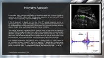
Innovative Approach Echographic scans are performed by EchoS device equipped with a convex transducer operating at 3.5 MHz, allowing the simultaneous acquisition of conventional B-mode images and corresponding unprocessed RF signals. R.E.M.S. approach is based on the idea that RF signals, acquired during an echographic scan of a target bone district, can be used to determine the health status of the considered bone through advanced comparisons with previously derived reference spectral models of the possible pathological or normal conditions. This method is natively integrated with US imaging,...
Open the catalog to page 6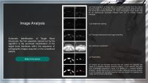
(a,b) Rearrangement of image data in rectangular matrices from the original sectorial images: (a) echographic image sample containing a vertebral interface that will be automatically identified, (b) "noisy" echographic image sample that will be automatically discarded because there are no suitable vertebral interfaces. Image Analysis (e,f) Contrast enhancement and image smoothing Automatic Identification of Target Bone Structures: the first operation carried out by the algorithm is the automatic identification of the target bone interfaces within the sequence of echographic images acquired on...
Open the catalog to page 7
Spectral Comparison Diagnostic parameter calculations are actually performed on RF signal segments corresponding to specific ROIs, which are automatically selected taking into account the position of the identified bone interfaces within the B-mode image. The principle behind is that the spectra of “raw” RF signals backscattered from the bone interface during an “in-vivo” echographic scan contain useful information about the bone status, including both quantity (e.g. BMD) and quality (e.g. elasticity) parameters. Accordingly, the O.S. value measures the degree of similarity between RF spectra...
Open the catalog to page 8
Osteoporosis Score Once all the frames belonging to the US dataset of the patient have been analyzed, the system verifies if the number of detected interfaces is sufficient to obtain a reliable diagnosis and calculations are performed on each RF spectrum of the identified ROIs. If this minimal frame condition is not satisfied (it happens rarely), the dataset is labeled as ‘‘noisy’’ and the operator is asked to repeat the US scan. For the O.S. calculation, the considered RF spectrum is classified as ‘‘osteoporotic’’ if the value of its Pearson correlation coefficient with the appropriate osteoporotic...
Open the catalog to page 9
Fragility Score* An analogous procedure is simultaneously applied to calculate the F.S.* value, with the only difference being that “osteoporotic” and “healthy” reference models are replaced by the corresponding spectral models of “frail” and “non-frail” bone structures. An important feature to be highlighted is the extreme ease of use of the system: inexperienced operators, who previously received a 3-hours specific training session only, performed acquisitions of a quality level appropriate for diagnostic calculations in 96.3% of cases (i.e. the incidence of “noisy” datasets was limited to...
Open the catalog to page 10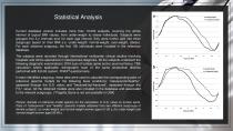
Statistical Analysis Current database version includes more than 10.000 subjects, covering the whole interval of typical BMI values, from under-weight to obese individuals. Subjects were grouped into 5-y intervals and, for each age interval, they were further split into three subgroups based on their BMI (i.e. under-weight/ normal-weight, over-weight, obese). For each obtained subgroup, the first 100 individuals were included in the reference database. The subjects were recruited through international multicenter clinical studies involving hospitals and clinics specialized in osteoporosis diagnosis....
Open the catalog to page 11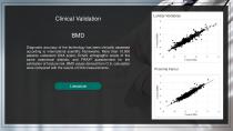
Clinical Validation Lumbar Vertebrae BMD Diagnostic accuracy of the technology has been clinically assessed according to international scientific frameworks. More than 10.000 patients underwent DXA scans, EchoS echographic scans of the same anatomical districts, and FRAX® questionnaire for the estimation of fracture risk. BMD values derived from O.S. calculation were compared with the results of DXA measurements. Proximal Femur
Open the catalog to page 12Archived catalogs
 REFERENCE SITES
21 Pages
 PRODCUTS
20 Pages