- Catalogs
- Erler-Zimmer
- 3D Anatomy Series
3D Anatomy Series
1 /9Pages

3D Anatomy Series
1 /9Pages
Catalog excerpts

Detailed article information at: www.erler-zimmer.de
Open the catalog to page 1
THE GROUND-BREAKING Monash Anatomy Series represents an unique and unrivalled collection of colour-augmented human anatomy body replicas designed specifically for enhanced teaching and learning. This premium collection of highly accurate normal human anatomy has been generated directly from either radiographic data or actual cadaveric specimens using advanced imaging techniques. The Monash 3D Human Anatomy Series provides a cost effective means to meet your specific educational and demonstration needs in a range of curricula from medicine, allied health sciences and biological sciences. A detailed...
Open the catalog to page 2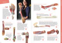
HAND/ARM Upper Limb – elbow, forearm and hand This 3D printed specimen demonstrates a superficial dissection of a left hand and wrist. Anteriorly, the transverse carpal and palmar carpal ligaments have been removed to expose the tendons and nerves traversing the carpal tunnel and Canal of Guyon. The palmar aponeurosis has been removed to demonstrate the course of the tendons through the palm, the superficial muscles of the thenar and hypothenar eminences (abductors and flexors), and the lumbrical Ref.no. MP1530 Details: muscles arising from the flexor digitorum tendon. Forearm and hand – superficial...
Open the catalog to page 3
Foot – Plantar surface Shoulder – deep dissection of the left shoulder joint, musculature, and associated nerves and vessels. This 3D printed specimen presents a deep dissection of the left shoulder joint, musculature, and associated nerves and vessels of the scapula and proximal humerus (to near midshaft). Anteriorly, the deltoid muscle has been detached from its origin to Ref.no. MP1525 Details: e xpose the underlying deeper s tructures of the shoulder joint and rotator cuff musculature. This 3D printed specimen provides a view of deep plantar structures of a right foot. Medially, the cut edge...
Open the catalog to page 4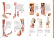
This 3D printed specimen consists of a right partial lower limb sectioned just proximal to the knee joint and complete through a partially dissected foot exposing the structures on the dorsum. Ref.no. MP1809 Details: Lower Limb superficial veins Popliteal Fossa distal thigh and This 3D printed specimen presents a superficial dissection of a left lower limb, from just proximal to the knee joint to a complete foot. The skin and superficial fascia have been removed to display the superficial venous structures of the leg including the dorsal venous plexus, great saphenous vein (including numerous...
Open the catalog to page 5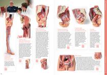
3D Printed Anatomy This 3D printed specimen presents a deep dissection of a left pelvis and thigh to show the course of the femoral artery and sciatic nerve from their proximal origins to the midshaft of the femur. Proximally, the pelvis has been sectioned along the mid-sagittal plane and the pelvic viscera are removed. In the pelvis the coccygeus muscle spans between the sacrum and iliac spine and the obturator artery and nerve entering the obturator canal superior to the obturator membrane. This 3D printed male left pelvis and proximal thigh (sectioned through the midsagittal plane in the midline...
Open the catalog to page 6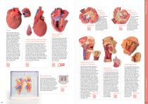
Heart internal structures This 3D printed specimen preserves the external anatomy of the heart and the distal trachea, carina, and primary bronchi in the posterior mediastinum relative to the great vessels and left atrium The left auricle has been sectioned to demonstrate the course of the circumflex artery in the coronary groove. The pulmonary trunk has been removed to expose the (open) pulmonary semilunar valves, while the arch of the aorta is intact to display the origins of the brachiocephalic trunk, left common carotid, and left subclavian. This 3D printed heart has been dissected to display...
Open the catalog to page 7
Superior Orbit This 3D printed model captures a dissection in which the calvaria and cerebrum have been removed to expose the floors of the anterior and middle cranial fossae. The midbrain has been sectioned at the level of the tentorium cerebelli and on the cross sectional surface one can identify the superior colliculi, cerebral peduncles and the substantia nigra. Anterior to the mid-brain the vertebral artery can be clearly identified rising Ref.no. MP1675 Details: from the posterior cranial fossa and dividing into the posterior cerebral arteries. Set of 3 Temporal Bone Model Medial Orbit...
Open the catalog to page 8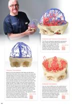
Arterial and Venous Circulation This 3D print integrates segmented angiographic data of both the cranial arterial and venous circulation into a single model. This Ref.no. MP1640 model is a combination of ‘Circle Details: of Willis’ MP1600, ‘Cranial Arterial Circulation’ MP1650 and ‘Cranial Venous Circulation’ MP1645 prints. Venous Circulation Based on the same dataset as MP1600 and MP1650, in this 3D print the dural venous sinus network has been segmented based on structures visible from the circulation of contrast medium. As a result, while most of the sinuses are present, the lack of contrast...
Open the catalog to page 9All Erler-Zimmer catalogs and technical brochures
 3D Anatomy Series
45 Pages
 Anatomical Charts
15 Pages
 Veterinary Medicine
44 Pages
 Anatomical Models
40 Pages
 Medical Simulators
84 Pages
Archived catalogs
 Catalog 2021
360 Pages
 X-Ray Phantoms
24 Pages

- Entrhal anatomical model
- Entrhal training anatomical model
- Entrhal teaching anatomical model
- Entrhal training simulator
- Positioning pillow
- Entrhal general care simulator
- Surgical anatomy model
- Entrhal test phantom
- Dental model
- Entrhal upper body simulator
- Entrhal flexible anatomical model
- Dental care model
- Entrhal skull model
- Entrhal training manikin
- Surgical phantom
- Transparent anatomical model
- Plastic anatomical model
- External defibrillator
- Mouth anatomical model