- Catalogs
- Erler-Zimmer
- 3D Anatomy Series
3D Anatomy Series
1 /45Pages
3D Anatomy Series
1 /45Pages
Catalog excerpts

Product Portfolio ERLER ZIMMER
Open the catalog to page 1
Exclusive 3D-printed anatomical specimens of the highest quality! Our premium collection of human body replicas was created directly from radiological data or real specimens using the latest imaging techniques. Human Body Replicas to Enhance Teaching! The groundbreaking anatomy series by Erler-Zimmer features a unique and unmatched collection of colored human body replicas, specifically designed to enhance teaching and learning. This premium collection of highly accurate human anatomy was created directly from radiological data or real specimens using the latest imaging technologies. The 3D Human...
Open the catalog to page 2
The ground-breaking Monash Anatomy Series represents an unique and unrivalled collection of colour-augmented human anatomy body replicas designed specifically for enhanced teaching and learning. Advantages of The Monash 3D Anatomy Series over plastic models or plastinated specimens: Realistic anatomical detail: Each replica is created from highresolution medical imaging or carefully selected real cadaver dissections. Unlike stylized plastic models, these represent real human anatomy with clinically important detail and high fidelity. Scientifically validated: All models are thoroughly reviewed...
Open the catalog to page 3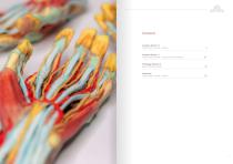
Content Anatomy Series 1.0 Didactically colored - healthy 8 Didactically colored - extended Series healthy 34 Rare Pathology cases 48 Didactically colored - healthy 80
Open the catalog to page 4
3D Anatomy Series | Anatomy Series 1.0
Open the catalog to page 5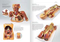
3D Anatomy Series | Anatomy Series 1.0 Head, Neck and Shoulder with angiosomes Nervous System Dissection (posterior view) This large 3D printed specimen displays a great deal of anatomy spanning the head, neck, thorax, axillae and upper limbs. This 3D printed specimen presents a unique view of axial anatomy, presenting a dorsal deep dissection of the head, neck, axillae, thorax, abdomen, and gluteal regions. The removal of the posterior portions of the cranium and laminectomy from the cervical region to the opening of the sacral canal affords a continuous view of the central nervous system structures...
Open the catalog to page 6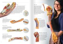
3D Anatomy Series | Anatomy Series 1.0 Hand This 3D printed specimen demonstrates a superficial dissection of a left hand and wrist. Anteriorly, the transverse carpal and palmar carpal ligaments have been removed to expose the tendons and nerves traversing the carpal tunnel and Canal of Guyon. The palmar aponeurosis has been removed to demonstrate the course of the tendons through the palm, the superficial muscles of the thenar and hypothenar eminences (abductors and flexors), and the lumbrical muscles arising from the flexor digitorum tendon. Item No. MP1530 Upper Limb This 3D-printed specimen...
Open the catalog to page 7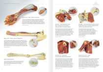
3D Anatomy Series | Anatomy Series 1.0 Upper Limb – elbow, forearm and hand This 3D-printed specimen displays a great deal of upper limb anatomy. In the distal arm and elbow/cubital fossa region it shows the arrangement of the biceps tendon, brachial artery and median nerve arranged from lateral to medial. The bicipital aponeurosis has been divided to reveal the structures deep to it. Item No. MP1510 Shoulder – deep dissection of the left shoulder joint, musculature, and associated nerves and vessels. Shoulder – deep dissection of a right shoulder girdle, preserving a complete scapula, lateral...
Open the catalog to page 8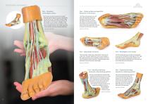
3D Anatomy Series | Anatomy Series 1.0 Foot – Structures of the plantar surface This 3D print records the anatomy of a right distal leg and the deep structures of the plantar surface of the foot. Proximally, the tibia, fibula, interosseous membrane, and leg muscles are discernable in cross-section. Medially, at the level of the ankle joint, the long tendons of the dorsi- and plantar-flexors are visible superficial to the c apsular and extra capsular ligaments. Item No. MP1900 Foot – Plantar surface and superficial dissection on the dorsum. This 3D printed specimen is a left foot with superficial...
Open the catalog to page 9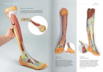
3D Anatomy Series | Anatomy Series 1.0 Lower Limb – deep dissection This 3D printed specimen consists of a right partial lower limb sectioned just proximal to the knee joint and complete through a partially dissected foot exposing the structures on the dorsum. Item No. MP1809 Lower Limb superficial veins Lower limb superficial dissection This 3D-printed specimen shows a superficial dissection of the left lower limb, from just above the knee to the foot. The skin and superficial fascia have been removed to reveal superficial veins, including the dorsal venous plexus, great saphenous vein with...
Open the catalog to page 10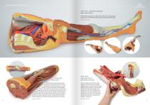
3D Anatomy Series | Anatomy Series 1.0 Lower limb – superficial dissection with male left pelvis This 3D printed specimen combines the Lower limb – superficial dissection (Ref.no. MP1816) with the male left pelvis (Ref.no. MP1765). Item No. MP1818 Lower Limb Musculature Lower Limb – deep dissection of a left pelvis and thigh This 3D printed specimen preserves a superficial dissection of the lower limb musculature from the mid-thigh to mid-leg, as well as nerves and vessels of the popliteal fossa. The insertions of the muscles of the anterior, middle and posterior compartments of the thigh are...
Open the catalog to page 11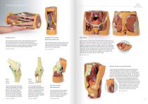
3D Anatomy Series | Anatomy Series 1.0 Popliteal Fossa This 3D-printed specimen shows the distal thigh and proximal leg, dissected posteriorly to reveal the popliteal fossa and surrounding structures. The proximal cross-section displays the compartment muscles, femoral vessels in the adductor canal, as well as the sciatic nerve and great saphenous vein. Item No. MP1830 Popliteal Fossa distal thigh and proximal leg This 3D printed specimen preserves the distal thigh and proximal leg, dissected posteriorly to demonstrate the contents of the popliteal fossa and surrounding region. Item No. MP1820...
Open the catalog to page 12
3D Anatomy Series | Anatomy Series 1.0 Heart This 3D printed heart specimen preserves superficial cardiac anatomy and the bases of the great vessels. All four chambers (atria and ventricles) are preserved, with the pericardial reflections on the left atrium demarcating the position of the transverse and oblique pericardial sinuses. On the posterior aspect, the coronary sinus receives all the cardiac veins (great, middle, small) and a prominent posterior vein of the left ventricle. The aortic and pulmonary semilunar valves are visible at the bases of the ascending aorta and pulmonary trunk, respectively....
Open the catalog to page 13All Erler-Zimmer catalogs and technical brochures
 Anatomical Charts
15 Pages
 Veterinary Medicine
44 Pages
 Anatomical Models
40 Pages
 Medical Simulators
84 Pages
Archived catalogs
 Catalog 2021
360 Pages
 3D Anatomy Series
9 Pages
 X-Ray Phantoms
24 Pages

- Entrhal anatomical model
- Entrhal training anatomical model
- Entrhal teaching anatomical model
- Entrhal training simulator
- Positioning pillow
- Entrhal general care simulator
- Surgical anatomy model
- Entrhal test phantom
- Dental model
- Entrhal upper body simulator
- Entrhal flexible anatomical model
- Entrhal bone model
- Dental care model
- Entrhal skull model
- Entrhal training manikin
- Surgical phantom
- Transparent anatomical model
- Plastic anatomical model
- External defibrillator
- Mouth anatomical model