- Company
- Products
- Catalogs
- News & Trends
- Exhibitions
G-scan Open
1 /20Pages

G-scan Open
1 /20Pages
Catalog excerpts
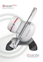
G•scanOPEN Beyond conventional MRI
Open the catalog to page 1
A revolutionary MRI approach The G-scan Open offers a groundbreaking scanning experience tailored for comprehensive musculoskeletal and spine imaging, enhancing diagnostic precision and confidence. A proprietary tilting design pushes the boundaries of traditional imaging by providing a unique perspective for positioning patients in both the supine and upright weightbearing positions, signaling a new era in imaging excellence. The G-scan Open is an ideal solution for spine surgeons, radiologist s , chiropractors , spor t s me dicine , and multidisciplinary clinics to acquire comprehensive assessments....
Open the catalog to page 2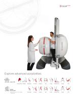
Explore advanced possibilities ** Supine MRI Hand/Wrist Elbow Weight Bearing MRI *Depending on room height **Optional
Open the catalog to page 3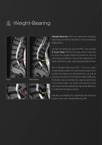
Weight-Bearing Weight-Bearing MRI is an advanced imaging technique primed to transform musculoskeletal diagnostics. Unlike conventional supine MRIs, the upright G-scan Open MRI technology allows patients to assume weight-bearing positions during scanning, providing a true-to-life assessment of their condition under natural physiological load. With Weight- Bearing MRI, clinicians gain unparalleled insight into spinal pathologies such as disc herniation and spinal stenosis, as well as precise evaluation of joint abnormalities affecting Supine the knees, hips, and ankles. By capturing the body in...
Open the catalog to page 4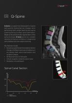
Q-Spine Q-Spine is a support tool developed to visualize and quantify biomechanical changes when performing both weight-bearing and supine scanning during a lumbar spine examination. Based on the semiautomatic segmentation of the spine structures, Q-Spine serves as a valuable resource to improve confidence in diagnosing spine diseases and pre-surgical planning. Key features include: • Numerical quantification that detects relative changes of different spine abnormalities such as Listhesis Index, spine canal section, and vertebral collapse. Virtual navigation inside the spinal canal. Spinal Canal...
Open the catalog to page 5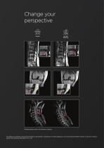
Change your perspective Supine Weight Bearing Weight-bearing exam with dynamic imaging *Courtesy of University L’Aquila and IOM Prof. Luigi Manfrè -Chairperson for Spine Diagnostic and Interventional ESNR, Director of the Mini-Invasive Spinal Interventional Operations Unit -IOM
Open the catalog to page 6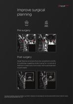
Improve surgical planning Supine Weight Bearing Post surgery Weight Bearing can be beneficial when evaluating the stability of a pathology, suggesting whether going for a conservative treatment or surgery and, in such a case, which surgical approach would fit better. *Courtesy of University L’Aquila and IOM Prof. Luigi Manfrè -Chairperson for Spine Diagnostic and Interventional ESNR, Director of the Mini-Invasive Spinal Interventional Operations Unit -I
Open the catalog to page 7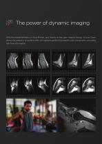
The power of dynamic imaging With the implementation of True-Motion, and thanks to the open magnet design, G-scan Open allows the operator to perform MRI with patients performing specific joint movements, providing real-time information.
Open the catalog to page 8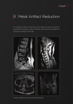
Metal Artifact Reduction Our advanced Metal Artifact Reduction sequence reduces distortion and artifacts caused by metallic implants, ensuring precise diagnostics and post treatment monitoring. * X-MAR is not available for sales in USA; MAR is available world-wide.
Open the catalog to page 9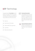
Technology Integrated inside the G-scan Open, the eXP Customized Examination platform provides advanced software With dedicated sequences, such acquisition techniques, such as Speed-Up as 3DHYCE for spine nerve roots, and TR reduction, to reduce scan time and improve diagnostic output and accuracy. Echo Dixon (SPED), the G-scan Open offers extensive protocols that can be customized for specific Speed-Up High resolution 3D isotropic X-Bone & SPED Dixon techniques True-Motion Metal Artifact Reduction Improved Diagnostic Experience Benefit from unique features, such as Metal Artifact Reduction and...
Open the catalog to page 10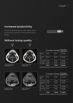
Increased productivity Scan more patients per day with Speed-Up, an acceleration acquisition method patented by Esaote. Without losing quality C-spine 3DHyce Axial Conventional 4’43” Conventional SpeedUp Exam time Reduction % C-spine 3DHyce Axial SpeedUp 2’57” Images and data results courtesy of University of L’Aquila, Italy Knee FSE T2 Axial Conventional 4’50” Knee FSE T2 Axial SpeedUp 3’44” Exam time Reduction %
Open the catalog to page 11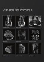
Knee SPED Coronal L-spine 3D HYCE Coronal Shoulder SPED Coronal
Open the catalog to page 12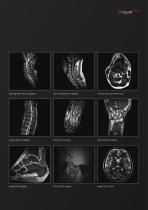
Cervical spine FSE T2 Sagittal Cervical spine SE T1 Sagittal Cervical spine 3D HYCE Axial Ankle SPED Sagittal
Open the catalog to page 13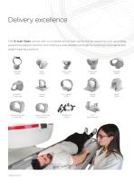
Delivery excellence The G-scan Open comes with a complete set of high-performance receiving coils, providing exceptional patient comfort, and covering a wide anatomical range for scanning in the supine and weight-bearing positions. Shoulder (linear) Cspine (linear) L-Spine, large size (4 channels) L-Spine, small size (4 channels) *Optional Coil
Open the catalog to page 14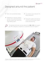
Designed around the patient Open, anti-claustrophobia design. Low ambient noise enhances the patient experience. Designed for maximum patient comfort with an optimized patient Wide gantry design and real-time bed that supports both supine and monitoring offer greater patient weight-bearing exams. “The premature termination rate was reduced from 58% to 8% when using an open MRI configuration compared to a conventional close-bore magnet”.** **Bangard C, Paszek J, Berg F, et al. MR imaging of claustrophobic patients inan open 1.0T scanner: motion artifacts and patient acceptability comparedwith...
Open the catalog to page 15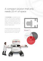
A compact solution that only needs 23 m2 of space The G-scan Open is a compact one-room solution, reducing upfront installation costs. This plug-and-play MRI system modern demand for healthcare as it combines low power consumption with represents a sustainable answer to the top level imaging quality. A dynamic stand-alone solution that offers a co m pl ete p atie nt eva l u atio n by performing both supine and weight- bearing examinations in a single scan. Dynamic assestment True-Motion
Open the catalog to page 16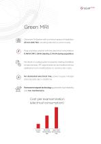
Green MRI One room Mri System with a minimum space of installation, 23 m2 (245 ft2), including electronics and console. Plug-and-play solution with low electrical consumption: 0.4kVA OFF, 1.2kVA stand by, 2.4 kVA during acquisition. No helium or cooling system is required, making installation simple and easy. RF cage shields can be installed without additional room modifications or construction costs. No dedicated electrical line; power supply voltage: 200-210/220-240 V 50/60 Hz Permanent magnet technology guarantees high reliability and low maintenance. Cost per examamination (electrical consumption)...
Open the catalog to page 17All Esaote catalogs and technical brochures
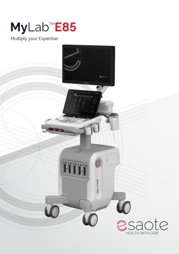 MyLab™E85
16 Pages
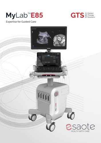 MyLab™E85 GTS
16 Pages
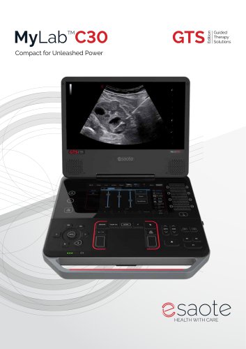 MyLab™C30 GTS
12 Pages
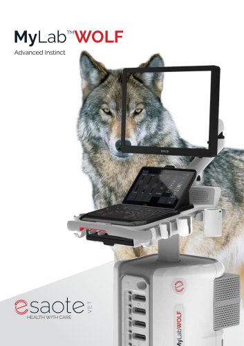 MyLab™Wolf
8 Pages
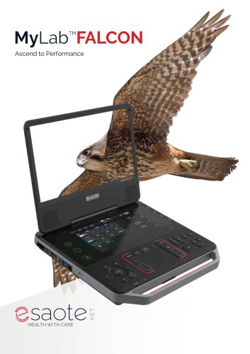 MyLab™Falcon
8 Pages
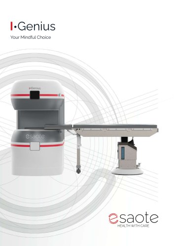 I-Genius
8 Pages
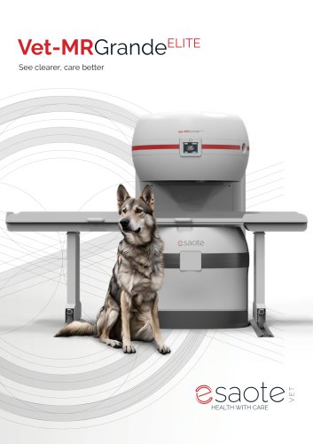 Vet-MR Grande ELITE
12 Pages
 MyLab™C30
12 Pages
 MyLab™C25
8 Pages
 MyLab™Heron
8 Pages
 Magnifico Open
16 Pages
 MyLab™X1 Go
4 Pages
 Q7
Q74 Pages
 MyLab™X1 Go VET
4 Pages
 Q7 VET
4 Pages
 O-scan VET
20 Pages
 Vet-MR Grande
8 Pages
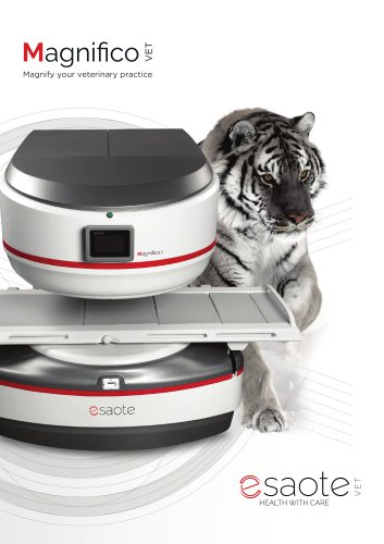 Magnifico Vet
12 Pages
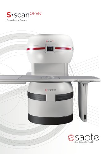 S-scan Open
12 Pages
 3MENSIO
13 Pages
 CAAS MR
2 Pages
 CAAS WORKSTATION
2 Pages
 MyLab™E80
9 Pages
 MyLab™Panther
12 Pages
 O-scan SMART
16 Pages
 MyLab™Fox
5 Pages
 MyLab™Omega eXP VET
12 Pages
 G-Scan Equine
8 Pages
 MyLab™A70
7 Pages
 MyLab™A50
5 Pages
 MyLab™X1
4 Pages
 MyLab™X90VET
8 Pages
 MyLab™Omega eXP CV Edition
12 Pages
 MyLab™Omega eXP
12 Pages
 MyLab™X90
24 Pages
 MyLab™X75VET
4 Pages
 MyLab™X1VET
4 Pages
 O-scan Premium & Light
12 Pages
 AgileExam
4 Pages
 MyLab™TringaVET
2 Pages
 MyLab™X75
12 Pages
 MyLab™X9
16 Pages
 MyLab™X8VET
2 Pages
 MyLab™9VET
2 Pages
 MyLab™X7VET
2 Pages
 MyLab™X5VET
2 Pages
 MyLab™OmegaVET
2 Pages
 MyLab™SigmaVET
2 Pages
 MyLab™VET Product Line
12 Pages
 MyLab™X8 eXP
12 Pages
 MyLab™X8 Platform
8 Pages
 MyLab™Omega
8 Pages
 MyLab™Sigma
4 Pages
 MyLab™X6
5 Pages
 MyLab™X5
5 Pages
 MyLab™X7
7 Pages
 MyLab™9 Platform
16 Pages
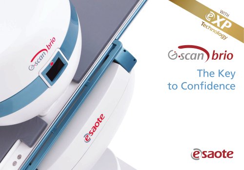 G-scan Brio 0.25 T
20 Pages
 S-scan
12 Pages


- Esaote ultrasound system
- Analysis software
- Esaote B/W ultrasound system
- Esaote color doppler ultrasound system
- Esaote portable ultrasound system
- Esaote multipurpose ultrasound imaging system
- Esaote visualization software
- Esaote medical imaging software
- Esaote convex-array ultrasound system
- Reporting software
- Esaote linear-array ultrasound system
- Esaote ultrasound transducer
- Esaote veterinary ultrasound system
- Esaote multipurpose veterinary ultrasound system
- Esaote on-platform ultrasound system
- Esaote portable veterinary ultrasound system
- Diagnostic software
- Hand-held ultrasound system
- Wireless probe ultrasound system
- Esaote touchscreen ultrasound system