


- Company
- Products
- Catalogs
- News & Trends
- Exhibitions
APX100

APX100
The APX100 Digital Imaging System by Olympus is designed to enhance research workflows by providing high-quality microscope images efficiently. It combines advanced optics, an intuitive interface, AI capabilities, and smart features to improve research productivity.
- Automatic Sample Detection: Utilizes a smart sample navigator and AI to automatically detect and center samples, allowing researchers to focus on image acquisition.
- High-Speed Autofocus: Offers faster autofocus than conventional systems, reducing time spent on sample searching.
- Gradient Contrast Method: Provides sharper images with higher contrast, suitable for both glass and plastic containers.
- Motorized Spherical Aberration Correction: Automatically adjusts for cover glass thickness to ensure clear images.
- Photobleaching Prevention Mode: Controls fluorescence illumination to minimize phototoxicity.
- Efficient Organization: Automatically organizes and indexes image data for easy management.
- Recall Settings: Saves acquisition settings with image data for easy replication in future experiments.
- TruSight Deconvolution: Enhances image quality by improving signal-to-noise ratio and resolution.
- cellSens Image Analysis: Provides advanced image processing techniques and deep-learning technology for efficient analysis.
- Flexible Imaging: Supports various imaging methods such as multichannel, stitching, time-lapse, and Z-stack acquisition.
- Wide Range of Objectives: Compatible with multiple Olympus objectives, including high-end options for diverse research needs.
- Includes various objectives, mirror units, and optional high-sensitivity cameras for enhanced imaging capabilities.
The APX100 microscope is available in two configurations: the standard unit (APX100-SU) and the high-end camera unit (APX100-HCU). It supports observation methods including brightfield, fluorescence, phase contrast, and gradient contrast. The sample holder accommodates glass slides, 35 mm dishes, and microplates. Users can choose from 25 objectives ranging from 4X to 100X, with a six-position motorized revolving nosepiece and motorized aberration correction.
The microscope features built-in Köhler illumination with high color rendering LED for transmitted light. It includes a motorized XY stage and focusing system. The fluorescence system includes a fly-eye lens, 18 mirror cubes, and an eight-position motorized mirror turret. A high brightness light guide and fluorescence LED illuminator are available, along with a motorized ND filter changer.
The system includes a 6.41-megapixel color and monochrome CMOS camera, with an optional high-sensitivity monochrome camera. The operation software is cellSens APEX, with optional software for time-lapse, wellplate navigation, full count and measure, and CI deconvolution.
Optional units include the Tokai Hit STX series and KSX-Type2. The system weighs approximately 34.6 kg to 35.3 kg and consumes 70 W of power. It operates on a power supply of AC 90–264 V, 50–60 Hz, with an output of DC 24 V/5 A.
The microscope requires Microsoft Windows 10 or 11 Pro (64-bit), a CPU equivalent to Intel Core processor (3.3GHz, 6 core), 16 GB RAM, and a display resolution of 1920 × 1080.
Evident Corporation is ISO 14001 and ISO 9001 certified. All specifications and appearances are subject to change without notice.
Catalog excerpts

Life Science APX100 Digital Imaging System Exceptional Imaging Made Easy
Open the catalog to page 1
Faster, Smarter Research Workflow The APEXVIEW APX100 digital imaging system makes it fast and simple to acquire expert-quality microscope images to make your research more efficient. Built with renowned Olympus optics, an intuitive user interface, a powerful AI, and a suite of smart features, the APX100 system combines the ease of use of an all-in-one-microscope with high-quality image data to fit your research needs. Find location Image acquisition Data management Time saved
Open the catalog to page 2
► Stay Focused on Your Research ► Publication-Quality Images in a Few Clicks ► Fast, Efficient Data Management
Open the catalog to page 3
Stay Focused on Your Research Spend More Time on Your Research with Automatic Sample Detection When you place your sample in the sample holder, the APX100 system’s smart sample navigator automatically acquires a macro image, and the built-in AI locates your sample on the slide. Once the system automatically centers your sample over the objective, it is displayed on the monitor so that you can choose the observation method and immediately begin capturing images. Click insert sample Step 1. Open the lid and place samples. The system accommodates dishes, slides, well plates, flasks, and more. Step...
Open the catalog to page 4
Simple Layout and Workflow Maximize Efficiency Using the APX100 system is simple—load the sample, close the lid, and click a button. The software’s clear layout and streamlined workflow help you start image acquisition with little training. Live View Window: For basic operations such as switching objectives, observation methods, and live acquisition. Stage Navigator: Control XY movement and stitching settings Macro Image: Display the entire holder Well Navigator: Image acquisition settings with microplates Process Manager: Multi-dimensional acquisition settings Camera Control: Various camera...
Open the catalog to page 5
Publication-Quality Images in a Few Clicks With an optical system designed and built by Olympus, the APX100 system delivers the same quality optics and technology available in our high-end research microscopes. This means that you can easily acquire publication-quality images for a wide range of applications. Mouse kidney. Alexa Fluor 488 WGA, Alexa Fluor 568 Phalloidin, DAPI. Rabbit liver. Stain: HE. Uncompromising Optical Technology Many Olympus objectives—including our award-winning X Line and silicone oil immersion objectives— work with the APX100 system, providing the flexibility to get...
Open the catalog to page 6
Mouse kidney. Alexa Fluor 488 WGA, Alexa Fluor 568 Phalloidin, DAPI. 7
Open the catalog to page 7
Gradient Contrast: See Your Sample in a New Light Acquiring sharp images at the edges of a well in a multi-well plate can be challenging. The contrast is poor even when using oblique contrast, and DIC does not work well with plastic containers. The APX100 microscope features our unique gradient contrast method. It works with all Olympus objectives and enables you to capture sharp images in a wider area and with higher contrast than the conventional method. This unique contrasting method is less affected by the meniscus, container lids, and water droplets, and it can be used with both glass- and...
Open the catalog to page 8
Sample Images Captured with Gradient Contrast Mouse kidney Expression of membrane -translocated mCherry in HEK293T cell* * mage data courtesy of Rie Saba, Ph.D., Division of Developmental Biology and Anatomy, Department of Anatomy, Kyoto I Prefectural University of Medicine.
Open the catalog to page 9
Tools for Clearer Images - Photobleaching Prevention Mode C onsistently bright incident light can cause photobleaching and phototoxicity that quickly degrades fluorescent samples. Using the system’s photobleaching prevention mode, the fluorescence illumination source only emits excitation light for the time necessary to acquire the image. The static image is then displayed on the screen, providing you time to take measurements or evaluate captures while minimizing phototoxicity to delicate samples. Live / snap acquisition Stage movement Illumination - A Line 20X Objective for Plastic Containers...
Open the catalog to page 10
Fast, Efficient Data Management Managing experiment data is a common challenge when acquiring multiple types of images across experiments. The APX100 system includes features that enable fast, efficient data management that keeps your data organized and acquisition parameters saved for future reference. Images Are Organized and Easy to Find The APX100 microscope has a dedicated system to organize and store your data. When you acquire an image, the software automatically creates folders for each sample and saves the data into the correct one. The consistent indexing keeps your data organized and...
Open the catalog to page 11
Image Processing and Analysis TruSight Deconvolution Fluorescence blur is a challenge when imaging thick specimens with widefield fluorescent microscopes. The APX100 features several tools for improving image quality, including online-deblur and TruSight deconvolution. The microscope’s TruSight deconvolution algorithms improve image quality, enabling you to obtain a higher signal-to-noise ratio, enhance resolution, and see deeper in thick specimens. When used in combination with a silicone oil immersion objective—which enables high-resolution and deep observation of cells and tissues—it can be...
Open the catalog to page 12
Designed for Labs and Imaging Core Facilities No Darkroom Required With a small footprint, built-in anti-vibration mechanism, and shielded optics, you can place the APX100 system almost anywhere. Install the unit directly on a lab table to image in parallel with other experiments, even in a brightly lit room. Easy to Learn, Easy to Teach Teaching multiple imaging modalities to new researchers can be a time-consuming process, but the APX100 system’s ease of use greatly simplifies and accelerates the training process. Researchers who are unfamiliar with microscopes can quickly learn to operate...
Open the catalog to page 13
Flexible for Many Applications The APX100 microscope supports a wide range of research imaging applications for slides, dishes, and well plates. Use the included imaging methods such as multichannel, stitching, time-lapse, and Z-stack acquisition in any combination to fit your research protocol. Multichannel • bserve samples with multiple stains and in combination with other imaging modes like phase contrast or O gradient contrast. • Capacity of up to eight mirror cubes to accommodate many experimental conditions. • apture images under optimal conditions quickly with automated exposure time and...
Open the catalog to page 14All Evident - Olympus Scientific Solutions catalogs and technical brochures
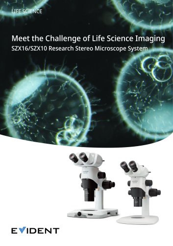 Stereo Microscope SZX16
24 Pages
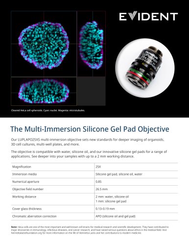 LUPLAPO25XS Silicone Gel Pad
2 Pages
 FV5000 / FV5000MPE
20 Pages
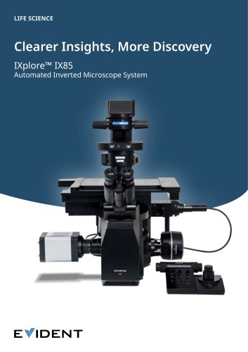 IXplore IX85 Pro
12 Pages
 DP23
DP238 Pages
 NoviSight brochure
4 Pages
 CellSens brochure
4 Pages
 X Line
2 Pages
 DP23M
DP23M4 Pages
 DP23/ DP23M/ DP28/ DP75/ SC180
12 Pages
 DP28
DP288 Pages
 VS200 brochure
12 Pages
 ScanR
ScanR8 Pages
 ICSI/IMSI brochure
8 Pages
 SZ61- SZ51
12 Pages
 SZX10, SZX16 family brochure
24 Pages
 SZX7
SZX716 Pages
 BX63/ BX53
8 Pages
 BX53- BX43- BX46 brochure
12 Pages
 CX3 Series
8 Pages
 CX23
CX234 Pages
 LC35
LC354 Pages
 CM30
CM308 Pages
 DP75
DP758 Pages
 FV4000MPE
4 Pages
 FV4000
20 Pages


- EVIDENT optical microscope
- EVIDENT laboratory microscope
- EVIDENT benchtop microscope
- EVIDENT LED microscope
- Camera with USB port
- Medical camera
- LED camera
- EVIDENT biology microscope
- Full HD camera
- HD camera
- EVIDENT microscope camera
- EVIDENT trinocular microscope
- Binocular microscope
- EVIDENT fluorescence microscope
- Digital microscope
- Teaching microscope
- EVIDENT achromatic microscope