- Company
- Products
- Catalogs
- News & Trends
- Exhibitions
FV4000
1 /20Pages

FV4000
1 /20Pages
Catalog excerpts

LIFE SCIENCE FLUOVIEW FV4000 Confocal Laser Scanning Microscope ... Transforming Precision Imaging - LIFE SCIENCE
Open the catalog to page 1
Empower Your Imaging Experiments Transform your images with the FLUOVIEW™ FV4000 confocal laser scanning microscope. Advanced imaging technology enables the acquisition of higher precision images, empowering researchers with more reliable data from their samples. With our breakthrough SilVIR™ detector at the core of the system, you can achieve much lower noise, higher sensitivity, and improved photon resolving capabilities. With the FV4000 confocal microscope, you can acquire higher-quality, quantitative image data in less time and with less effort. Experience the system’s innovations, including:...
Open the catalog to page 2
In 2022, Olympus spun off its scientific solutions division—including life science microscopy—into a new, independent company we call Evident. Although our name is different, our expertise, manufacturing capabilities, and commitment to our customers developed over the past 100 years remain unchanged. As an independent company, we can innovate faster and develop better products. We think that the FV4000 microscope exemplifies this, and we can’t wait for you to discover what it can do for yourself.
Open the catalog to page 3
Easy-to-Acquire, Quantitative Confocal Data The FV4000 microscope uses our advanced, silicon-based SilVIR™ detector that makes it easier than ever to acquire precise, reproducible data. Multicolor image of C. elegans hybrid strain of NeuroPAL strain and GCaMP strain. NeuroPAL strain was generated by Eviatar Yemini and Oliver Hobert. Courtesy of Kotaro Kimura; Graduate School of Science, Nagoya City University and Asuka Takeishi; Neural circuit of multisensory integration, RIKEN Hakubi Research Team. Tip of a Drosophila leg (42-hour pupation), stained with phalloidin (AlexaFluor 405, F-actin,...
Open the catalog to page 4
Game-Changing Quantification The technology behind our SilVIR detector enables you to precisely quantify image intensity for more reliable data. Imaging data can be displayed as to the number of photons, providing the absolute value of the fluorescence intensity for each image. The wider dynamic range provides accurate quantification of fluorescence intensity by photon number even at high intensity levels. Cos-7 cells: anti-Tubulin (Alexa Fluor 488; green). Sample Courtesy of: Dr. Jana Döhner, Dr. Urs Ziegler, University of Zürich. SilVIR Intensity: 2330 Photon number: 73 Intensity: 745 Photon...
Open the catalog to page 5
Experience the Full Dynamic Range of Fluorescence Instead of choosing to focus on either dim or bright fluorescence areas, the FV4000 microscope can capture both in one image without saturation or loss of information thanks to the SilVIR detector’s high dynamic range. This allows accurate image analysis and processing with less work. Intuitive User Interface and Workflows The photomultiplier tubes traditionally used in confocal imaging require voltage adjustments depending on the sample’s brightness level as well as an offset adjustment to reduce signal noise. This requires expert knowledge and...
Open the catalog to page 6
Reproducible Image Data Between Users and Systems The SilVIR detector has less sensitivity loss over time than previous-generation detector technologies. With our laser power monitor (LPM) and TruFocus™ Z-drift compensator, achieve reproducible images under consistent conditions for better reproducibility. Different users on different days can acquire the same precise images using the same settings. Even the images acquired by different FV4000 microscopes can be compared and discussed using the same photon number intensity scale. SilVIR Next-Generation Detector Technology The SilVIR detector...
Open the catalog to page 7
More Information from Your Images See Further with NIR Capabilities The system's enhanced technologies enable expanded multiplexing to see more in one image. Our updated TruSpectral™ technology combined with high-sensitivity SilVIR detectors enable you to multiplex up to six channels simultaneously. The upgraded spectral system is comprised of our highly efficient volume phase hologram (VPH) grating and slit and can detect an industry-leading 400 nm to 900 nm wavelength range1 with a minimum step of 1 nm. Add up to six channels with your choice of broadband and red-shifted detectors. This setup...
Open the catalog to page 8
Cytoskeleton sample: HeLa cells stained with DAPI (blue), Pericentrin (Centrosome, green), a-Tubulin (Microtubules, Alexa-568; red) and phalloidin (Actin, Alexa-647; magenta). Sample Courtesy of: Sample preparation Alexia Ferrand; sample acquisition Sara R. Roig and Alexia Ferrand. Imaging Core Facility, Biozentrum, University of Basel. The neurovascular unit of a mouse hippocampus. Cell nuclei (DAPI; blue), astrocytes(AF488 GFAP; green), pericytes (DsRed; yellow), basement membrane of blood vessels (AF647 collagen IV; magenta), Astrocytes water channel (AF750 AQ-4; gray)'. -SamDleSourtesy of:...
Open the catalog to page 9
Flexible Macro to Micro Imaging Fast, Efficient Multiscale Observation The macro-to-micro workflow enables you to easily observe the target sample from the macro level—whole body or tissue—down to the cellular or subcellular level. High Image Quality at High Speed The system’s unique combination of advanced technologies delivers high-quality images faster than conventional laser scanning microscope systems. The 1k × 1k resonant scanner at FN20 with 0.033 µs per pixel enables you to rapidly acquire high-resolution images with minimal noise using the SilVIR detector. The result is that you can...
Open the catalog to page 10
Muscle tissue, blue; DAPI, green; FDGFR, red; F4/80. Sample courtesy of: Marshall Hogarth and Jyoti Jaiswal, Center for Genetic Medicine Research, Children’s National Research Institute. A total of 77 four-channel XYZ positions (11 × 7) were acquired using a 1K resonant scanner within 16 minutes to create the stitched image, which used to require 2 hours using a galvanometer scanner. The coronal section of an H-line mouse brain, cyan; DAPI (cell nuclei), green; YFP (neuron), yellow; Cy3 astrocytes, magenta; AlexaFluor 750 (microtubule). Sample courtesy of: Takako Kogure and Atsushi Miyawaki,...
Open the catalog to page 11All Evident - Olympus Scientific Solutions catalogs and technical brochures
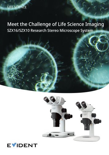 Stereo Microscope SZX16
24 Pages
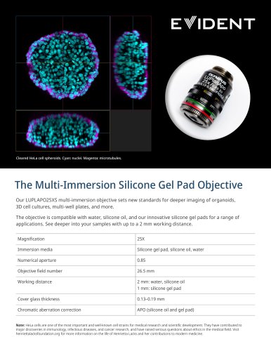 LUPLAPO25XS Silicone Gel Pad
2 Pages
 FV5000 / FV5000MPE
20 Pages
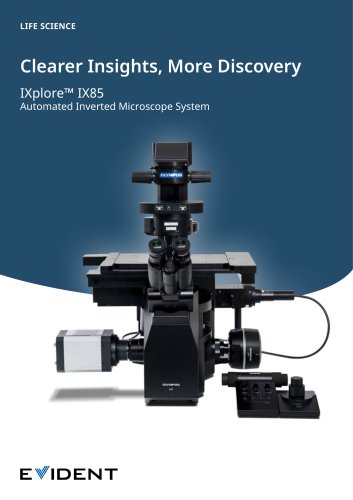 IXplore IX85 Pro
12 Pages
 DP23
DP238 Pages
 NoviSight brochure
4 Pages
 CellSens brochure
4 Pages
 X Line
2 Pages
 DP23M
DP23M4 Pages
 DP23/ DP23M/ DP28/ DP75/ SC180
12 Pages
 DP28
DP288 Pages
 VS200 brochure
12 Pages
 ScanR
ScanR8 Pages
 ICSI/IMSI brochure
8 Pages
 SZ61- SZ51
12 Pages
 SZX10, SZX16 family brochure
24 Pages
 SZX7
SZX716 Pages
 BX63/ BX53
8 Pages
 BX53- BX43- BX46 brochure
12 Pages
 CX3 Series
8 Pages
 CX23
CX234 Pages
 LC35
LC354 Pages
 CM30
CM308 Pages
 APX100
20 Pages
 DP75
DP758 Pages
 FV4000MPE
4 Pages


- Koko optical microscope
- Koko laboratory microscope
- Koko benchtop microscope
- Koko LED microscope
- Koko CMOS camera
- Camera with USB port
- Medical camera
- Koko biology microscope
- LED camera
- Full HD camera
- Koko trinocular microscope
- Koko microscope camera
- HD camera
- Binocular microscope
- Koko fluorescence microscope
- Koko research microscope
- Digital microscope
- Teaching microscope
- Koko achromatic microscope