- Catalogs
- Evident - Olympus Scientific Solutions
- VS200 brochure
- Company
- Products
- Catalogs
- News & Trends
- Exhibitions
VS200 brochure
1 /12Pages

VS200 brochure
1 /12Pages
Catalog excerpts

LIFE SCIENCE SLIDEVIEW VS200 The Power to See More
Open the catalog to page 1
Reliable Data for Many Applications Digitizing slide data makes it easy to analyze, share, and archive your results. The SLIDEVIEW VS200 research slide scanner enables you to capture high-resolution images of your slides for quantitative analysis, so you can make the most of the information your slides have to offer. The optical system is optimized for scanning slides, enabling you to digitize slides for brain, cancer, and stem cell research, as well as drug discovery. Brain Research Brain and neuroscience researchers need to observe various samples in detail—from single cells to entire tissue,...
Open the catalog to page 2
Multiplex Scan Mode When tissue samples are limited, it is critical to gather the most data possible from each tissue section. Multiplexing immunofluorescence allows for greater understanding of co-expression and the spatial composition of multiple targets within a single sample. The multiplexing scan mode helps optimize the utility of these select samples by aligning multiple fluorescent channels with a reference channel. Lung tissue imaged on a VS200 at 20x stained with an Ultivue PD-L1 kit multiplex kit; Dapi: Nuclear Counterstain, FITC: CD8, TRITC: CD68, Cy5: PD-L1, Cy7: panCK. Image data...
Open the catalog to page 3
Outstanding Image Quality for Quantification Better Resolution and Flatness To produce high-quality virtual slide images, the VS200 system uses X Line high-performance objectives, which offer simultaneously improved numerical aperture, chromatic aberration correction, and flatness. The result is flatter images with a wider field of view and negligible intensity fall off near the periphery. To further enhance the image quality, the system’s light path is optimized to work with X Line objectives, providing more homogenous illumination. These enhancements allow for excellent image quality so that...
Open the catalog to page 4
Achieve More in Less Time High Throughput The loader holds up to 210 26 × 76 mm (1 × 3 in.) slides in 35 slide trays. The robotics in the loader move the trays and not the individual slides, helping your slides remain safe and intact. The type of slide trays, number of slides, and the size of the slides are immediately detected, while the integrated barcode reader automatically captures and records the slide information. Higher Productivity Work on scan parameter settings for some slides while other slides are being acquired. The convenient software gives you the flexibility to control all of...
Open the catalog to page 5
Flexible for Many Applications Five Observation Methods in One System The VS200 slide scanner can be used for brightfield, fluorescence, darkfield, phase contrast, and simple polarization. This flexibility allows you to combine different observation methods to view structures that are only visible under certain conditions. For example, darkfield helps to get a proper overview image of a fluorescence sample unstained in the visible spectrum and provides the best contrast scaling between the overview signal and focused fluorescence signal. Phase contrast Human cartilage captured with X Line UPLXAPO10X...
Open the catalog to page 6
Supports Glass Slides and Plates The simple-to-use slide tray supports 26 × 76 mm (1 × 3 in.), 52 × 76 mm (2 × 3 in.), 76 × 102 mm (3 × 4 in.), and 102 × 127 mm (4 × 5 in.) slides. The system enables you to manage different slide sizes at the same time in the same batch scan. Flexibility to Use Dry, Silicone Oil, or Oil Objectives Unlike many slide scanners that do not offer highmagnification capabilities, the VS200 system's automatic oil dispenser enables you to use high-magnification, oil, or silicone oil immersion objectives for batch scanning without having to frequently stop to oil the lens....
Open the catalog to page 7
High-Contrast Optical Sectioning of Whole Slide Scans The Speckle Illumination Acquisition (SILA) optical sectioning device uses laser speckles to obtain highcontrast images by removing out-of-focus light. The HiLo microscope technology used by the device— developed by Bliq Photonics—offers many benefits and can easily be added to existing VS200 slide scanners. Widefield (left) and SILA (right) images of a whole planarian flatworm Schmidtea mediterranea in 20x, showing the intestines. Blue: DAPI. Green: inner intestine cells; Red: outer intestine cells. Samples provided by Amrutha Palavalli,...
Open the catalog to page 8
Deep Learning for Deeper Insights TruAI technology uses deep learning to simplify workflows and rapidly deliver more accurate results. Conventional thresholding methods often have difficulty identifying morphologic features on a sample and can miss critical targets. With a trained neural network, for example, on pancreatic samples, TruAI technology can accurately segment pancreatic islets and differentiate them from similar looking clusters of erythrocytes, enabling the number and size of the islets to be counted and measured automatically. (a) Cy3 fluorescence marked pancreatic islets. Pancreatic...
Open the catalog to page 9
Seamless from Scanning to Sharing Managing the large amount of data generated by your VS200 scanner is easier than ever thanks to compatibility with our NIS-SQL database and OlyVIA web programs. You can automatically upload your images to one or more databases, differentiate between users, and take advantage of offline visualization and annotation tools. Supports the of DICOM Format For clinical researchers working in a lab, the VS200 scanner enables you to save images in the DICOM format and upload them directly into your organization’s picture archiving and communication system (PACS) and connect...
Open the catalog to page 10
Specifications VS200 Single Tray Intended Specimen VS200 Multiple Tray Loader Observable Specimen Glass slide with cover glass Size of Glass Slide (W × L × H) Standard slide tray: 25 mm–26.5 mm × 75 mm–76.5 mm × 0.9 mm–1.2 mm (1 in. × 3 in. × 0.05 in.) (6 slides) Optional trays 1) 51 mm–53 mm × 75 mm–76.5 mm × 0.9 mm–1.2 mm (2 in. × 3 in. × 0.05 in.) (3 slides) 2) 100 mm–102 mm × 75 mm–76.5 mm × 0.9 mm–1.2 mm (4 in. × 3 in. × 0.05 in.) (1 slide) 3)126 mm–128 mm × 75 mm–76.5 mm × 1.1 mm–1.4 mm (5 in. × 3 in. × 0.06 in.) (1 slide) Cover Glass Thickness Observation Methods Brightfield, reflected...
Open the catalog to page 11All Evident - Olympus Scientific Solutions catalogs and technical brochures
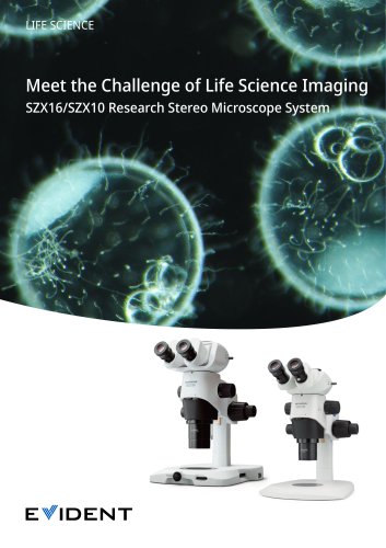 Stereo Microscope SZX16
24 Pages
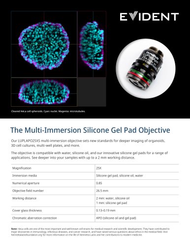 LUPLAPO25XS Silicone Gel Pad
2 Pages
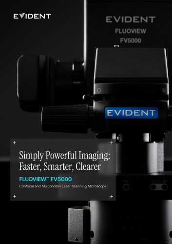 FV5000 / FV5000MPE
20 Pages
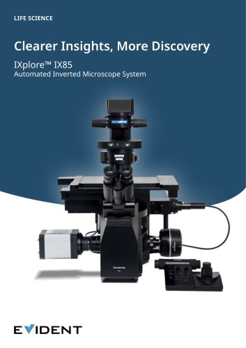 IXplore IX85 Pro
12 Pages
 DP23
DP238 Pages
 NoviSight brochure
4 Pages
 CellSens brochure
4 Pages
 X Line
2 Pages
 DP23M
DP23M4 Pages
 DP23/ DP23M/ DP28/ DP75/ SC180
12 Pages
 DP28
DP288 Pages
 ScanR
ScanR8 Pages
 ICSI/IMSI brochure
8 Pages
 SZ61- SZ51
12 Pages
 SZX10, SZX16 family brochure
24 Pages
 SZX7
SZX716 Pages
 BX63/ BX53
8 Pages
 BX53- BX43- BX46 brochure
12 Pages
 CX3 Series
8 Pages
 CX23
CX234 Pages
 LC35
LC354 Pages
 CM30
CM308 Pages
 APX100
20 Pages
 DP75
DP758 Pages
 FV4000MPE
4 Pages
 FV4000
20 Pages


- Koko optical microscope
- Koko laboratory microscope
- Koko benchtop microscope
- Koko LED microscope
- Koko CMOS camera
- Camera with USB port
- Medical camera
- Koko biology microscope
- LED camera
- Full HD camera
- Koko trinocular microscope
- Koko microscope camera
- HD camera
- Binocular microscope
- Koko fluorescence microscope
- Koko research microscope
- Digital microscope
- Teaching microscope
- Koko achromatic microscope