- Catalogs
- GE Healthcare Life Sciences
- Cytell™ Cell Imaging System
Cytell™ Cell Imaging System
1 /8Pages

Cytell™ Cell Imaging System
1 /8Pages
Catalog excerpts

GE Healthcare Life Sciences Data file 29-0866-95 AA Cell analysis and imaging Cytell™ Cell Imaging System The Cytell Cell Imaging System (Fig 1) combines the functionalities of a digital microscope, an image cytometer, and a cell counter in a single benchtop instrument. This compact, application-driven, automated cell imaging system provides robust quantitative results through the use of preconfigured biological applications (BioApps). Each BioApp is an easy-to-use, automated module that covers all steps of a specific biological application or assay. Cytell Cell Imaging System comes bundled with five BioApps to simplify routine cell lab tasks while providing high-quality scientific data. Fig 1. Cytell Cell Imaging System. Flexibility • Cytell can be used as a digital microscope, image cytometer, and cell counter. This flexibility simplifies logistics in the laboratory, reducing both the amount of equipment required and the dependence on core imaging facilities and associated expertise. • Ready-to-use reagent kits are available from GE Healthcare for cell counting, cell cycle, and cell viability applications. Alternative reagents are also supported as long as they match the four available fluorescent channels. • Cytell works with multiwell plates, slides, Petri dishes, and flasks, so you can capture great images from a range of samples. Table 1. Examples of recommended fluorophores for each fluorescent channel • Images and numerical data are saved in standard formats that can be imported into a variety of image analysis and statistical data programs. • Four color channels allow you to multiplex up to four fluorescent markers; with spectral optics optimized for commonly used dyes such as Hoechst™, GFP, Cy™3, and Cy5 (Table 1). Example Dyes DAPI, Hoechst, Alexa Fluor™ 405 GFP, Fluorescein, Calcein AM, Alexa Fluor 488, Cy2 Cy3, Rhodamine, TRITC, Alexa Fluor 546, Propidium Iodite, DsRed
Open the catalog to page 1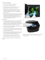
Hardware highlights • Out-of-the-box installation and operation • No preventive maintenance required • Solid-state illuminator: lifetime of 10 000 h, which is approximately 5× longer than a typical lamp • Automated XY stage, objective changer, Z-axis focus adjustment, emission filter changer, and autofocus; control all hardware components with user-adjustable imaging protocols • Sample motion, imaging operations, and other steps of the experimental procedure are performed either automatically or manually using the highly intuitive graphical user interface (GUI); automation simplifies instrument...
Open the catalog to page 2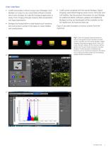
User interface • Cytell functionality is driven using a set of BioApps. Each BioApp is an easy-to-use, automated software module that covers all steps of a specific biological application or assay, from imaging through analysis, data visualization, and report generation. • BioApps are hosted within a web-based touch sensitive GUI environment, similar to the apps on newer tablets and smartphones. • Cytell comes complete with five starter BioApps: Digital Imaging, Automated Imaging, Quick Count, Cell Cycle, and Cell Viability. See the product information for each BioApp for additional details....
Open the catalog to page 3
Imaging performance Cytell is designed to capture high-quality cellular images suitable for collecting robust statistical measurements from subcellular features and cell populations. This capability is achieved by combining large field of view (FOV) and subcellular imaging resolution. Performance at low magnification: • Large FOV (2.2 × 1.6 mm) • Optimal for imaging whole cells, small organisms, and colonies • Several hundreds of cells can be captured in one image • Preview scan large areas to easily find an object of interest Some of these applications are shown in Figure 4. Performance at high...
Open the catalog to page 4
Data visualization Cytell provides a variety of interactive graphical tools to analyze and visualize experimental data from multiple wells, cell populations, and subpopulations. Five of the most commonly used tools, graphs, and plots are: • Histogram: examine population intensity distributions and apply gating thresholds to identify subpopulations; a histogram is an important tool for cell cycle phase analysis • Plate heat map: see at a glance how a single experimental parameter varies across the plate, for example in a dose-response experiment • Pie chart: visualize the relative size of cell...
Open the catalog to page 5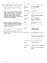
Results and other data files System specifications Each Cytell BioApp creates a number of data files to capture protocols, system configuration, and experimental results: Self-installed, out-of-the-box operation Instrument dimensions Input voltage Power requirements Imaging modes Epi-fluorescent and transmitted light wide field imaging Solid-state illuminator for fluorescence imaging LED for transmitted light imaging • Data files: Cell-by-cell and population measurements are saved in standard .csv format for easy import into thirdparty data analysis software such as Excel™ and FlowJo™. Fluorescence...
Open the catalog to page 6
Ordering information Products GE Cytell Cell Imaging System comprising instrument, workstation, microscope slide holder, 35 mm Petri dish holder, 60 mm Petri dish holder, 100 mm Petri dish holder, T-25 flask holder, T-75 flask holder, reference slide Code number Cytell Microscope Slide Holder Cytell 35 mm Petri Dish Holder Cytell 60 mm Petri Dish Holder Cytell 100 mm Petri Dish Holder Cytell T-25 Flask Holder Cytell T-75 Flask Holder Cytell Quick Count slides Related Reagents Code number Cytell Quick Count/Viability Reagent Cytell Cell Viability Kit Cytell Cell Viability Plus Reagent Cytell Cell...
Open the catalog to page 7
Products are for research purposes only. They are not approved for diagnosis of disease in humans or animals. GE, imagination at work, and GE monogram are trademarks of General Electric Company. Cy and Cytell are trademarks of GE Healthcare Companies. CyDye: This product is manufactured under an exclusive license from Carnegie Mellon University and is covered by US patent numbers 5,569,587 and 5,627,027. The purchase of CyDye products includes a limited license to use the CyDye products for internal research and development but not for any commercial purposes. A license to use the CyDye products...
Open the catalog to page 8All GE Healthcare Life Sciences catalogs and technical brochures
 ImageQuant TL 8.1
2 Pages
 Biacore T200
9 Pages
 Xcellerex XDUO Quad
4 Pages
 ÄKTA pilot 600
5 Pages


Archived catalogs
 Watman Syringe Filter Collection
26 Pages
 ÄKTA pure data file
12 Pages
 ÄKTAprocess
4 Pages
 ÄKTApurifier
12 Pages
 ÄKTAmicro
8 Pages
 ImageQuant LAS 500 data file
8 Pages
 Ultrospec 9000/9000PC
2 Pages
 Ultrospec 8000/8000PC
2 Pages
 Ultrospec 7000/7000PC
2 Pages
 WAVE Bioreactor 200 System
4 Pages
 The full imaging spectrum
32 Pages
 Typhoon Multislide Tray
2 Pages
 IN CELL ANALYZER 2200
12 Pages
 DeltaVision OMX
8 Pages
 Amersham Imager 600
8 Pages
 ImageMaster 2D Platinum
4 Pages
 SimpliNano spectrophotometer
4 Pages
 Xuri™. Believe in better futures
20 Pages
 ÄKTA avant
12 Pages


