- Catalogs
- GE Healthcare Life Sciences
- The full imaging spectrum
The full imaging spectrum
1 /32Pages

The full imaging spectrum
1 /32Pages
Catalog excerpts

The full imaging spectrum Superb images today. Powerful research tomorrow.
Open the catalog to page 1
Digital excellence in biomolecular imaging GE Healthcare Life Sciences offers a broad range of high-quality products covering 1-D and 2-D electrophoresis, Western blotting, and 2-D Difference Gel Electrophoresis (2-D DIGE). Common to all of these analytical workflows is image capture. Our portfolio of versatile and upgradeable imagers covers the entire range of protein and nucleic acid detection methods–from densitometry, through phosphorimaging to fluorescence and chemiluminescence–so that you can select the most appropriate analysis platform for your applications. Start-to-finish solutions...
Open the catalog to page 2
Chemiluminescence is the method of choice for sensitive, nonradioactive Western blotting. Amersham™ imagers provide quantitative imaging of chemiluminescence, and gel documentation. These CCD camera-based imagers provide publication grade data, digital image archiving, and better quantitation than film, without sacrificing sensitivity. Amersham Imager 600 QC is a dedicated configuration for densitometry applications in a QC environment. Fluorescence Fluorescence enables great sensitivity, as well as the detection of multiple samples in one gel or blot for accurate comparison. Amersham imagers...
Open the catalog to page 3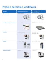
Protein detection workflows Versatile quantitative protein detection Chemiluminescence only Transfer, markers, 2nd dimension Rainbow markers TC 70/77 PWR Semi-dry Transfer Unit Amersham ECL Amersham ECL Select™ Amersham ECL Select Amersham ECL Prime Amersham ECL Plex™ Amersham Imager 600 ImageQuant LAS 500 Amersham Imager 600 UV Amersham Imager 600 RGB Extended anal
Open the catalog to page 4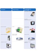
2-D electrophoresis and 2-D DIGE Ettan™ IPGphor™ 3 IEF System Ettan DALTsix Large Electrophoresis System Silver Stain Coomasie CyDye™ DIGE Dyes Amersham ECL Plex ImageMaster™ 2D Platinum DeCyder
Open the catalog to page 5
Typhoon laser scanners At the cutting edge of quantitative imaging The ability to distinguish subtle differences in expression among low amounts of biomolecules in complex mixtures adds power to many areas of biological research. Highly versatile Typhoon laser scanners enable you to generate data of the highest quality through linearity of signal response, quantitative accuracy, and extremely low limits of detection. Typhoon scanners utilize the strengths of laser-excited fluorescence and storage phosphor technology to achieve optimal signal detection for your samples. The imaging of radiolabels...
Open the catalog to page 6
Uncompromising scanning power Typhoon FLA 9500 supports phosphorimaging, 2-D DIGE imaging, red/green/blue (RGB) and near IR fluorescence as well as sensitive and accurate quantitation of proteins using the multifluorescent Amersham ECL Plex Western blotting system. High resolution and quantitation • Precisely quantitate signals across five orders of magnitude in gels, blots, tissue sections, and arrays up to 40 × 46 cm • Measure minute changes in expression by detecting signals otherwise obscured by background noise, down to 10 μm pixel resolution Versatility • Image multifluorescent, colorimetric/gel...
Open the catalog to page 8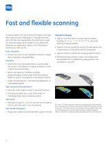
Fast and flexible scanning If a typical week in your lab involves the analysis of one blot after another by your colleagues on a shared instrument, each with their own requirements, the last thing you need is to lose time preparing the imager for your experiment. Whatever your application, Typhoon FLA 7000 scanner shortens your start-up time. Multi-user power • Shorten start-up time with predefined methods, multiple lasers and easily changeable filters Versatility • Choose from four preinstalled lasers to quickly adapt the system to the detection of signals emitted by a wide variety of fluorescent...
Open the catalog to page 10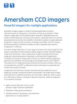
Amersham CCD imagers Powerful imagers for multiple applications Amersham imagers support a range of imaging applications including chemiluminescence, fluorescence, colorimetry, and gel documentation. These charge-coupled device (CCD)-based systems provide an affordable option for protein and DNA detection and reduce the need for a dedicated dark room and the associated environmental costs. Benefits of Amersham systems include publication grade data, image and data archiving, and better quantitation than film without sacrificing sensitivity. Amersham imagers are fully compatible with powerful...
Open the catalog to page 12
Great images are a touch away Plug-in start Getting started couldn’t be easier – to install the system, simply connect to the power source and switch on. The CCD is cooled and ready to deliver sensitive detection in just 5 minutes. • The optimized light path and a sensitive 8.3 megapixel cooled CCD are specifically designed for chemiluminescence Touchscreen A very simple touchscreen interface controls all imaging tasks. And naturally, it can be operated wearing lab gloves. Simple loading Loading trays are provided to image blots and gels. Simply place the sample on the relevant tray and insert...
Open the catalog to page 13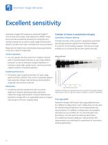
Amersham imager 600 series Excellent sensitivity Amersham imager 600 employs an advanced Fujifilm™ CCD camera and a bright, wide-aperture FUJINON™ F0.85 lens to provide exceptional sensitivity for Amersham ECL chemiluminescence, as well as a wide range of fluorescent labels for protein and nucleic acid detection and quantitation. Responses are highly linear and enable precise quantitation across four orders of magnitude. Freedom of choice in quantitative imaging Quantitative Western blotting The high sensitivity of the system is designed to quantitate the chemiluminescent signals from Amersham...
Open the catalog to page 14
Amersham imager 600 series Multiplexed images on a single membrane Amersham Imager 600 RGB is designed for multichannel imaging and is especially suited to the quantitation of signals on blots processed using Amersham ECL Plex Western blotting system with Cy™3 and Cy5. Independent visualization and quantitation of proteins completely removes the need for stripping and re-probing the membrane. A wide range of visible fluorescent dyes can be imaged via RGB epi-illumination combined with appropriate filter sets. UV fluorescence A UV transilluminator is available for fluorescence detection of reagents...
Open the catalog to page 16All GE Healthcare Life Sciences catalogs and technical brochures
 ImageQuant TL 8.1
2 Pages
 Biacore T200
9 Pages
 Xcellerex XDUO Quad
4 Pages
 ÄKTA pilot 600
5 Pages
 Cytell™ Cell Imaging System
8 Pages


Archived catalogs
 Watman Syringe Filter Collection
26 Pages
 ÄKTA pure data file
12 Pages
 ÄKTAprocess
4 Pages
 ÄKTApurifier
12 Pages
 ÄKTAmicro
8 Pages
 ImageQuant LAS 500 data file
8 Pages
 Ultrospec 9000/9000PC
2 Pages
 Ultrospec 8000/8000PC
2 Pages
 Ultrospec 7000/7000PC
2 Pages
 WAVE Bioreactor 200 System
4 Pages
 Typhoon Multislide Tray
2 Pages
 IN CELL ANALYZER 2200
12 Pages
 DeltaVision OMX
8 Pages
 Amersham Imager 600
8 Pages
 ImageMaster 2D Platinum
4 Pages
 SimpliNano spectrophotometer
4 Pages
 Xuri™. Believe in better futures
20 Pages
 ÄKTA avant
12 Pages


- Solvent reagent
- Molecular biology reagent
- Analysis software
- Protein reagent
- Histology reagent
- Sterile needle
- Dye reagent kit
- Cytology reagent
- Control software
- Buffer solution reagent
- Medium reagent
- Antibody
- PCR reagent
- Bacteria reagent
- Virus reagent
- Automated software
- Serum reagent
- DNA extraction reagent
- Reagent for nucleic acids
- Laboratory filter