
- Catalogs
- GE Healthcare
- Discovery MR750


- Products
- Catalogs
- News & Trends
- Exhibitions
Discovery MR750

Discovery MR750
This technical document appears to be related to medical imaging, specifically Magnetic Resonance Imaging (MRI) protocols and sequences for various anatomical regions and clinical applications. The content includes references to imaging parameters, anatomical focus areas, and imaging techniques.
1. Imaging Modalities and Techniques
- SignaPULSE and Related Sequences: Mentioned as part of the imaging protocols, likely referring to GE Healthcare MRI pulse sequences.
- Water only, Fat only, In-phase, Out-of-phase Imaging: Techniques used to differentiate tissue types based on their chemical composition, important for fat suppression and characterization.
- Multi-station Spine Imaging: Protocols for imaging the entire spine in multiple stations to cover cervical, thoracic, and lumbar regions.
2. Anatomical Focus Areas
- Musculoskeletal Imaging: Protocols and parameters for imaging bones, joints, and soft tissues.
- Abdominal and Vascular Imaging: Techniques for imaging abdominal organs and vascular structures, including fat suppression and contrast techniques.
- Neuro Imaging: Brain and spinal cord imaging protocols, emphasizing depiction of spinal cord and disc spaces.
- Cardiac Imaging: Sequences and parameters tailored for heart imaging, likely including functional and structural assessment.
- Spine Imaging: Detailed protocols for axial cervical spine imaging, highlighting excellent depiction of spinal cord and intervertebral disc spaces.
3. Imaging Parameters and Protocols
- Repeated references to parameters such as TR (repetition time), TE (echo time), and specific sequence codes (e.g., 8340; 550; 570) suggest detailed protocol settings for different anatomical regions.
- Use of fat suppression techniques and phase-sensitive imaging to improve tissue contrast.
- Multi-planar imaging and multi-station approaches to cover extended anatomical regions.
4. Recommendations and Best Practices
- Emphasis on excellent depiction of critical structures such as spinal cord and disc spaces, indicating the importance of high-resolution imaging and appropriate sequence selection.
- Use of multi-station imaging for comprehensive spine assessment.
- Application of chemical shift imaging techniques (water/fat separation) to enhance diagnostic accuracy.
Catalog excerpts
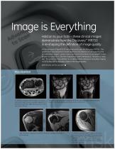
Image is Everything Hold on to your hats - these clinical images demonstrate how the Discovery™ MR750 is re-shaping the definition of image quality. In the last issue of SignaPULSE we introduced to you the Discovery MR750 - the system that was designed to break traditional boundaries of 3.0T scanning. We provided white papers, system specs, and technical articles so you wouldn't have to take our word for it. And the response has been tremendous. However, as they say, "the proof is in the pudding" so, to demonstrate what your everyday imaging could be like, we've included a taste of the image quality. We'll let you see for yourself. ■ 3D MERGE acquisition with 60 slices 2 mm thick Small FOV, high resolution imaging of the wrist trabeculae and carpometacarpal scanned through the wrist. Excellent depiction of joints. 1 mm acquired slices thickness, 1024 x 1024 matrix. all tendons with 288 x 288 matrix and 12 cm FOV. Scan time 2:34 min. High-resolution imaging of the glenoid, articular cartilage, and rotator cuff structures.
Open the catalog to page 1
LAVA-Flex study in pelvis displaying 4 unique images acquired at each slice location. A total of 84 images scanned in a 19 s breath hold with a 320 x 256 imaging matrix. A TR of 4.8 ms allows acquisition of two echoes at 1.1 and 2.2 ms which correspond to the out and in phase images (at 3.0T). The water and fat only images are subsequently reconstructed from the acquired 1.1 and 2.2 ms TE images with perfect separation of the 1H in fat and water. The result is exquisite suppression of fat (or water) which enables excellent visualization of pathology.
Open the catalog to page 2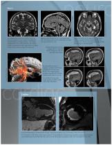
T2 weighted Fast Spin Echo PROPELLER 2.O in the coronal plane is very useful to visualize the temporal lobes and hippocampus. 3 mm slices imaged with a 22 cm FOV and 512 x 512 matrix results in a pixel size of 43O x 43O microns, 15 slices in Scan Time 2:45 min using 32 ch brain coil. iogimd HAIR i-fiCWLLLfi ^ * min . l D‘_ v. :S 530- JPJhhotrutv.-rlhm LlL 411 afawi &S iliCe n r) scan! nut of i l] rrvi •'lRfbil T hi:, image i:.\v-IS sl'Cft'j 5c.trnr:i w*IFi JCl lersar ;yi\"tcrj. ?WE-H ft- L 44 mil The -mining Tl itl-jglflthy rv£QC dipi^-L whrft irdltn ffcfn iSrknLtTuF- rlfl[AivveM£i?iprl rra1isnri-,Ji.lmt^^ill...
Open the catalog to page 3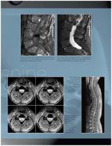
Multi-station spine scan with IDEAL exhibiting excellent fat exclusion along the entire length of the spine. 3D MERGE in the axial cervical spine. 60 slices covering 3 disc spaces in 2:50 min with a 288 x 256 matrix. Excellent depiction of the grey and white matter of the spinal cord.
Open the catalog to page 5All GE Healthcare catalogs and technical brochures
 Vscan Air™ CL
6 Pages
 Voluson™ Expert Series
4 Pages
 Vivid T9
9 Pages
 SIGNA™ Explorer
34 Pages
 Versana Active™
2 Pages
 GSI infographic
2 Pages
 MAC 2000 ECG Analysis System
5 Pages
 EMR Gateway Pro for MAC 2000
2 Pages
 Revolution EVO Gen 3
28 Pages
 Seno Iris™
5 Pages
 SWAN
SWAN2 Pages
 GE Adventure Series™
14 Pages
 Mobile X-ray Systems
10 Pages
 GoldSeal
8 Pages
 Invenia™ABUS
6 Pages
 CardioMem CM 3000
2 Pages
 Lullaby Resus Plus
4 Pages
 Brivo NM615
5 Pages
 Aespire 7900
6 Pages
 Aespire View
4 Pages
 Aisys Carestation
12 Pages
 Avance Carestation
12 Pages
 Lunar iDXA Brochure
8 Pages
 Brochure Discovery IGS 730
9 Pages
 Prodigy for Bone Health
8 Pages
 Senographe Care
7 Pages
 SenoBright White Paper
7 Pages
 SenoBright Brochure
20 Pages


- GE ultrasound system
- GE B/W ultrasound system
- GE color doppler ultrasound system
- Portable ultrasound system
- Multipurpose ultrasound imaging system
- Patient monitor
- Radiology software
- Visualization software
- Tablet computer software
- Tablet PC software
- Digital radiography system
- Reporting software
- Phototherapy lamp
- Convex-array ultrasound system
- Linear-array ultrasound system
- Blood pressure patient monitor
- Multipurpose radiography system
- X-ray system
- ECG patient monitor
- SpO2 patient monitor