- Catalogs
- GE Healthcare
- SenoBright White Paper
- Products
- Catalogs
- News & Trends
- Exhibitions
SenoBright White Paper

SenoBright White Paper
Breast cancer is a major health concern with a 12.5% lifetime risk for women. Imaging techniques such as mammography, ultrasound, and contrast-enhanced MRI (CE-MRI) are essential for screening and diagnosis. However, CE-MRI is often limited by cost and availability. GE Healthcare's SenoBright, introduced in 2010, offers a cost-effective alternative through Contrast Enhanced Spectral Mammography (CESM), which provides both morphological and contrast-enhanced images using an iodinated contrast agent and low radiation dose.
Contrast-Enhanced Spectral Mammography (CESM)
CESM uses dual-energy X-ray acquisitions to highlight hypervascularized breast lesions. It enhances iodine detectability by suppressing background breast tissue, using low-energy (LE) and high-energy (HE) X-ray spectra tailored for each breast thickness. The HE spectra are optimized with a multi-layer copper filter to maximize iodine contrast.
Average Glandular Dose (AGD)
SenoBright's AGD is only 20% higher than standard mammography, providing a diagnostic view at about half the maximum AGD limit set by the American College of Radiology, ensuring lower radiation exposure and enhanced patient safety.
SenoBright Solution
SenoBright integrates into existing GE mammography systems, focusing on high-quality imaging, low radiation exposure, and efficient workflow. The recombination algorithm processes LE and HE images to highlight iodine-enhanced areas, ensuring clear visualization of lesions.
Clinical Workflow and Patient Comfort
The SenoBright exam is designed for efficiency and patient comfort, taking less than ten minutes. It involves a single iodine injection and rapid image acquisition, allowing for immediate image review and streamlined workflow.
Clinical Summary
CESM has evolved from early X-ray imaging techniques, overcoming limitations of temporal subtraction methods. Recent studies demonstrate CESM's effectiveness in lesion localization and its potential as a problem-solving tool compared to standard mammography and ultrasound.
Conclusion
SenoBright enhances diagnostic capabilities by providing quick, high-quality imaging that highlights unusual blood flow in breast lesions. It allows for immediate follow-up tests using existing mammography equipment, improving patient care and workflow efficiency.
Case Study Overview
The document includes a case study of a patient diagnosed with invasive ductal carcinoma, confirmed by biopsy, with ultrasound imaging of the left breast.
References
The document cites various studies and guidelines related to breast cancer diagnosis and imaging techniques, including SEER Cancer Statistics Review, ACR practice guidelines for mammography, and research on dual-energy contrast-enhanced digital mammography.
Company Information
The document is published by General Electric Company, specifically GE Healthcare, which reserves the right to modify product specifications and features. Contact information and trademarks are provided.
Catalog excerpts
SenoBright Contrast Enhanced Spectral Mammography Technology Ann-Katherine Carton Sylvie Saab-Puong Matt Suminski White Paper October 2012
Open the catalog to page 1
SenoBright Contrast Enhanced Spectral Mammography Technology Ann-Katherine Carton Sylvie Saab-Puong Matt Suminski Introduction Women have up to a 1 in 8 (12.5%) lifetime risk of developing breast cancer.1 To improve health outcome, imaging techniques are key in screening, diagnosis and therapy for breast cancer. In a diagnostic setting today, mammography, ultrasound and contrast-enhanced MRI (CE-MRI) are the standard imaging tools to help define suspicious lesions previously seen in screening mammograms. CE-MRI is complementary to mammography and ultrasound since it provides additional functional...
Open the catalog to page 2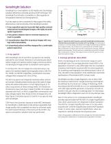
SenoBright is a novel addition to GE Healthcare’s technology solutions delivering a clinically practical CESM technique. The SenoBright functionality is available in a field upgrade on Senographe Essential and Senographe DS. Four key aspects were considered to help support the safety, effectiveness, and practicality of the SenoBright solution: 1. X-ray acquisition spectra to provide high-quality contrastenhanced and morphological images, with highly accurate spatial registration 2. A low patient radiation dose to minimize exposure as much as possible 3. A recombination algorithm to produce images...
Open the catalog to page 3
3. Recombination algorithm An optimized recombination algorithm on SenoBright is dedicated to process the LE and HE images into iodine images.3 The algorithm, using as input the LE and HE X-ray spectra and the compressed breast thickness, is designed to efficiently suppress the background breast tissue for all breast thicknesses and density patterns, in order to highlight the iodineenhanced areas. The recombination algorithm ensures the visibility of 0.5 mg/cm² iodine areal concentration, corresponding to a lower bound of concentration clinically expected. An iodine image computed with our exclusive...
Open the catalog to page 4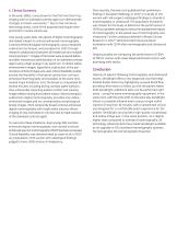
5. Clinical Summary In the early 1980s, it was shown for the first time that X-ray imaging with an iodinated contrast agent can demonstrate changes in breast vascularity.4,5 Due to the immature technology status, none of the proposed techniques were practical for routine clinical use. Only twenty years later, the advent of digital mammography stimulated interest in contrast-enhanced mammography. Contrast-enhanced digital mammography, using a temporal subtraction technique, was pioneered in 2002 through research collaborations between GE Healthcare and multiple clinical partners.6,7 Images of...
Open the catalog to page 5
SenoBright Case Study Figure 4a: Original mammography from a 79 y/o patient who presented with palpable mass on left breast. From left: Right cranial-caudal (RCC), Left Cranial-Caudal (LCC), Right Medial-Lateral Oblique (RMLO), and Left Medial-Lateral Oblique (LMLO). No particular findings, but very dense breast tissue. Figure 4b: Ultrasound of the left breast of same patient. Figure 4c: SenoBright Contrast Enhanced Spectral Mammography exam of the same patient. From left: LCC low-energy, LCC Contrast-enhanced, LMLO contrast-enhanced, LMLO low-energy. The contrast-enhanced images clearly localize...
Open the catalog to page 6
References 1. http://seer.cancer.gov/csr/1975_2009_pops09/results_merged/sect_04_ breast.pdf 2. LW Bassett et al., ACR practice guideline for the performance of screening and diagnostic mammography, 2008 3. S Puong et al., Dual-energy Contrast Enhanced Digital Mammography using a new approach for breast tissue canceling, Proc. SPIE Medical Imaging, vol. 6510, pp. 65102H, 2007 4. LV Ackerman et al. Breast lesions examined by digital angiography. Work in progress. Radiology vol 155, pp.65-68, 1985 5. AC Watt et al. Breast lesions: differential diagnosis using digital subtraction angiography. Radiology,...
Open the catalog to page 7All GE Healthcare catalogs and technical brochures
 Vscan Air™ CL
6 Pages
 Voluson™ Expert Series
4 Pages
 Vivid T9
9 Pages
 SIGNA™ Explorer
34 Pages
 Versana Active™
2 Pages
 GSI infographic
2 Pages
 MAC 2000 ECG Analysis System
5 Pages
 EMR Gateway Pro for MAC 2000
2 Pages
 Revolution EVO Gen 3
28 Pages
 Discovery MR750
5 Pages
 Seno Iris™
5 Pages
 SWAN
SWAN2 Pages
 GE Adventure Series™
14 Pages
 Mobile X-ray Systems
10 Pages
 GoldSeal
8 Pages
 Invenia™ABUS
6 Pages
 CardioMem CM 3000
2 Pages
 Lullaby Resus Plus
4 Pages
 Brivo NM615
5 Pages
 Aespire 7900
6 Pages
 Aespire View
4 Pages
 Aisys Carestation
12 Pages
 Avance Carestation
12 Pages
 Lunar iDXA Brochure
8 Pages
 Brochure Discovery IGS 730
9 Pages
 Prodigy for Bone Health
8 Pages
 Senographe Care
7 Pages
 SenoBright Brochure
20 Pages


- Capintec ultrasound system
- Capintec B/W ultrasound system
- Capintec color doppler ultrasound system
- Patient monitor
- Portable ultrasound system
- Multipurpose ultrasound imaging system
- Visualization software
- Radiology software
- Tablet computer software
- Tablet PC software
- Digital radiography system
- Reporting software
- Phototherapy lamp
- Convex-array ultrasound system
- Blood pressure patient monitor
- Linear-array ultrasound system
- ECG patient monitor
- Multipurpose radiography system
- X-ray system
- SpO2 patient monitor