- Catalogs
- Hamilton Thorne
- Oosight
Oosight
1 /4Pages

Oosight
1 /4Pages
Catalog excerpts

HAMILTON THORNE See what you've been missing. OpsTghf Imaging System ^^5 Adding Oosight® to your lab can improve success by giving you a quantitative and reproducible method to measure biological disruption in either fresh or previously frozen oocytes. You can now select oocytes for ICSI and embryos for implantation, and use the system to help improve enucleation efficiency. Understanding the oocyte is critical to understanding embryogenesis, and studies show that a disrupted spindle apparatus or a weakened zona pellucida in the oocyte can yield lower pregnancy rates. In fact, it has been shown that pregnancy is up to 8 times more likely when the inner zona pellucida is well-ordered.1 The unique and patented solid-state, liquid crystal technology is an easy add-on to your ICSI workstation. Oosight software runs on your computer to capture, display, and analyze your images. Snap an image and click a button to report the data. Meaningful data on molecular order within the sample are organized into an intuitive, exportable report. It's really that simple. Key Benefits • Unprecedented Resolution High-contrast live images of the oocyte and spindle • Non-invasive Imaging Does not require the use of any labels or stains, preserving the biology of the spindle and related structures • Quantitative Analysis Tracks oocyte behaviour over time and automatically records data points of molecular density and orientation • Proven Successfully used to image many different mammalian species for both enucleation and developmental studies The Oosight® Imaging System is for research purposes only. Shen Y, et. al. High magnitude of light retardation by the zona pellucida is associated with conception cycles. Human Reproduction, 2005 Jun; 20(6):1596-606.
Open the catalog to page 1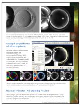
In a conventional contrast image (left) of a human MII oocyte taken just prior to ICSI, structures such as the spindle and multiple layers of the zona pellucida remain invisible. In an Oosight image (right) the spindle is clearly seen to be nicely barrel shaped and the three layers of the zona pellucida are all visible. Oosight outperforms all other systems. No other contrast-enhancement technique delivers the performance of Oosight. With unprecedented resolution and calibrated setup, Oosight provides the sensitivity and reproducibility required of a grading routine, as well as the speed needed...
Open the catalog to page 2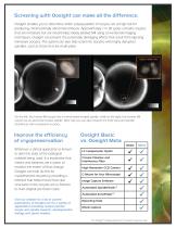
Screening with Oosight can make all the difference. Oosight enables you to determine which subpopulation of oocytes are at high risk for producing chromosomally abnormal embryos. Approximately 1 in 20 cycles contains oocytes that are immature but are nevertheless falsely labeled MII using conventional imaging techniques. Oosight can prevent the potentially damaging effects that result from injecting immature oocytes. The system can also help screen for oocytes with highly disrupted spindles, such as those that are multi-polar. On the left, this human MII oocyte has a normal barrel-shaped spindle,...
Open the catalog to page 3
All specifications subject to change. Optical Wavelength of operation Spatial resolution Electrical Power source 5V 3A with universal input voltage adapters Image Acquisition Image output format Scientific CCD Camera Sensor size Image size Pixel dimensions Digital output Binning modes Computer Requirements PC Desktop/Laptop Operating System Memory Hard disk Display USB ports Minimum Intel Penitum, 2 GHz Windows 7, 32-bit 1 GB 80 GB 1280 x 1024 USB 2.0, 2 available ports Microscope Compatibility Oosight systems are compatible with many research-grade microscopes, including those made by Leica®,...
Open the catalog to page 4All Hamilton Thorne catalogs and technical brochures
 Lykos & Zilos-tk
4 Pages
 VibeXactive
1 Page
 Vibex passive
1 Page
 CLINICAL LASER HARDWARE
1 Page
 STILETTO® RESEARCH LASER
1 Page
 Axio Vert.A1
14 Pages
 SteREO Discovery.V8
10 Pages
 Stemi 508
30 Pages
 Axio Lab.A1
18 Pages
 CT37stax Brochure
12 Pages

- Solvent reagent
- Research reagent
- Laboratory reagent
- Diagnostic reagent
- Laboratory incubator
- Dye reagent kit
- Quality control reagent
- Clinical reagent
- Benchtop laboratory incubator
- Stainless steel laboratory incubator
- Laboratory air sterilizer
- Micropipette
- Scientific research reagent
- Cell imaging system
- Automatic cell imaging system
- Laboratory cell imaging system
- Portable air sanitizer
- Laser module
- Continuous monitoring system
- Micromanipulator