- Catalogs
- Hangzhou Antigenne Technology Co. Ltd
- Instruction Manual-Vamber-C.T&S IgE(80)
- Company
- Products
- Catalogs
- News & Trends
- Exhibitions
Instruction Manual-Vamber-C.T&S IgE(80)
1 /2Pages
Instruction Manual-Vamber-C.T&S IgE(80)
1 /2Pages
Catalog excerpts
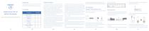
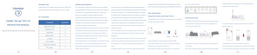
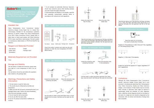
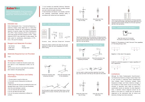
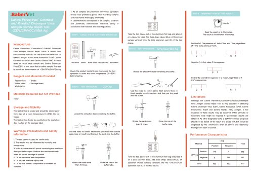
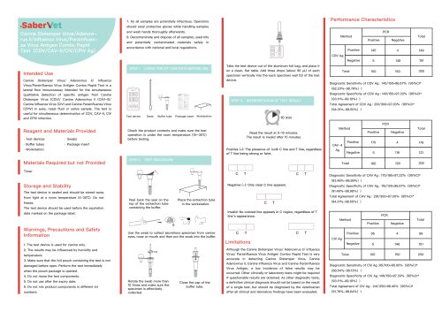
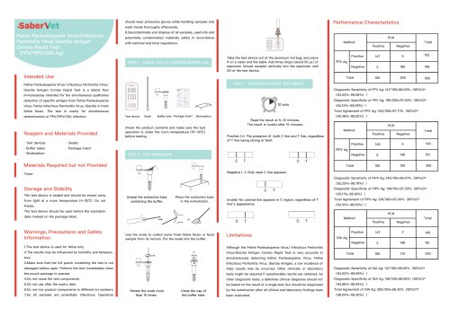
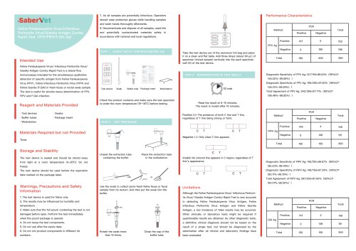
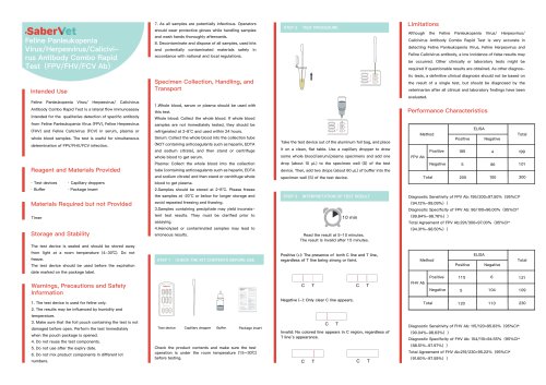
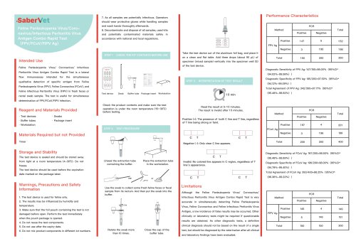
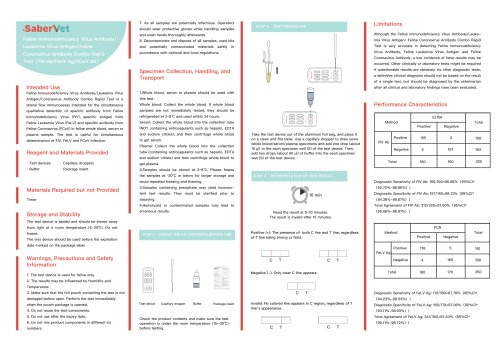
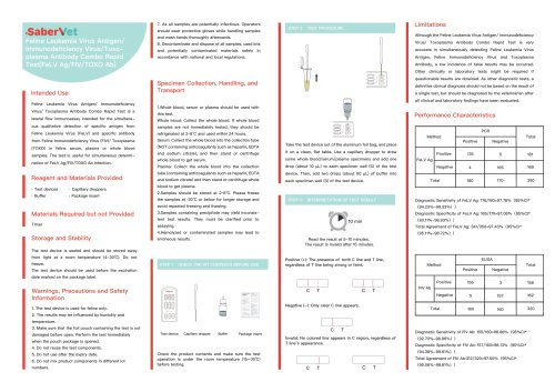
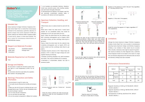
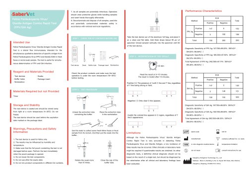
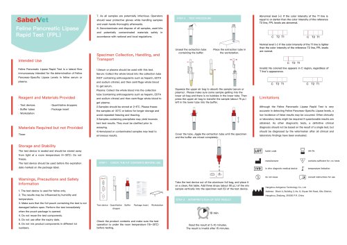
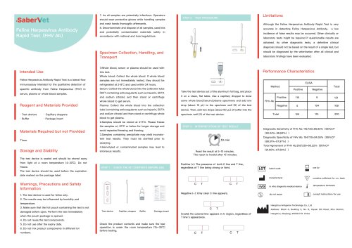
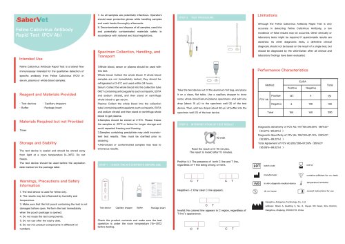
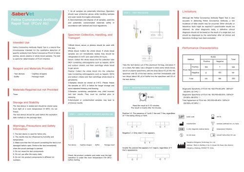
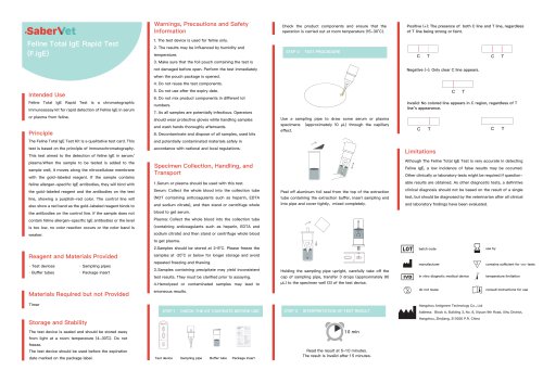
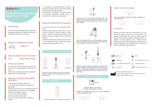
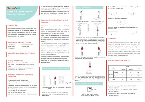
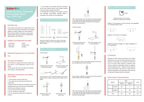
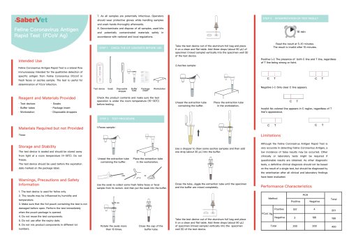
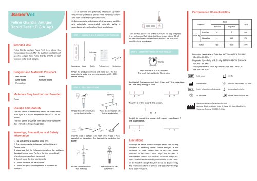
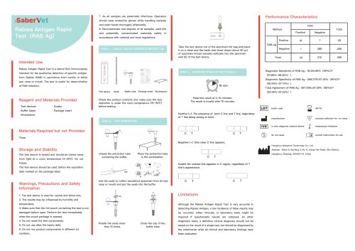
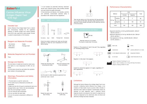
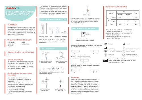
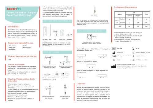
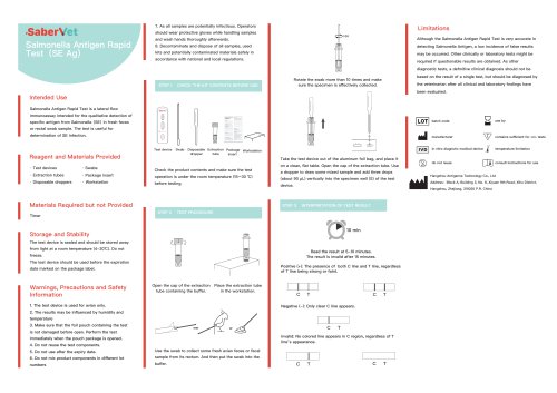
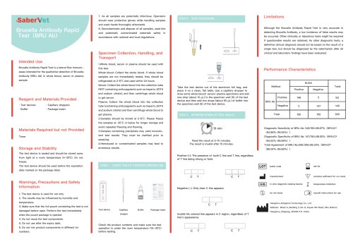
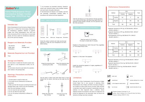
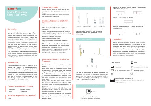
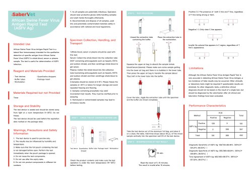
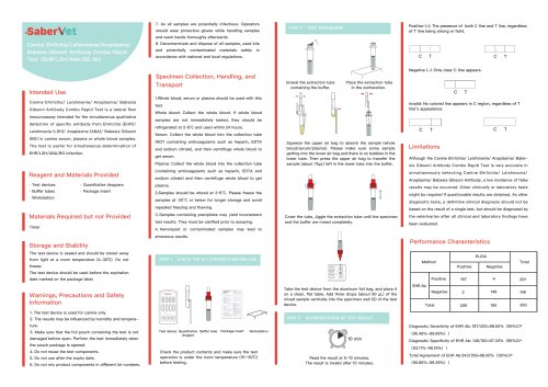
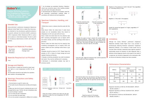
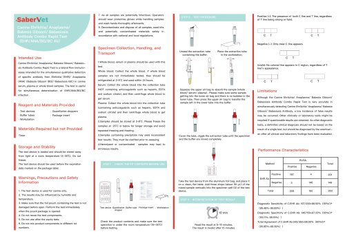
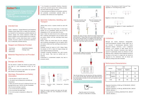
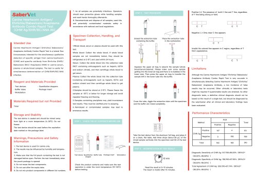
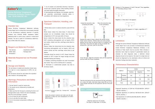
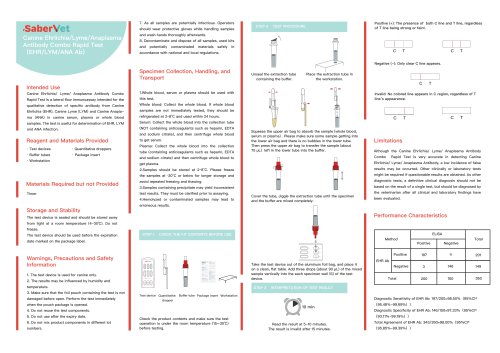
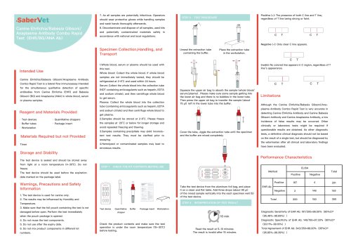
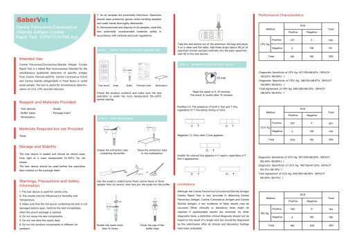
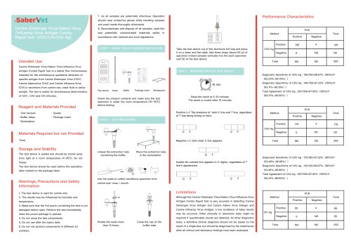
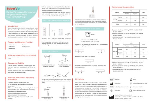
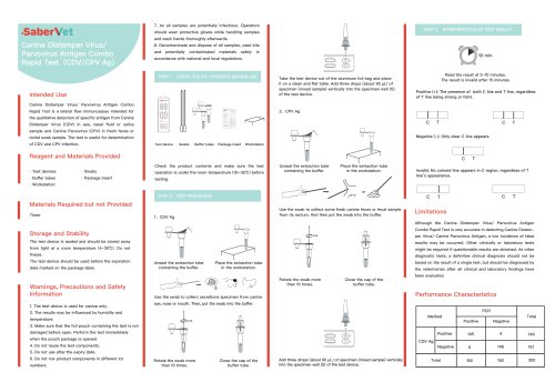
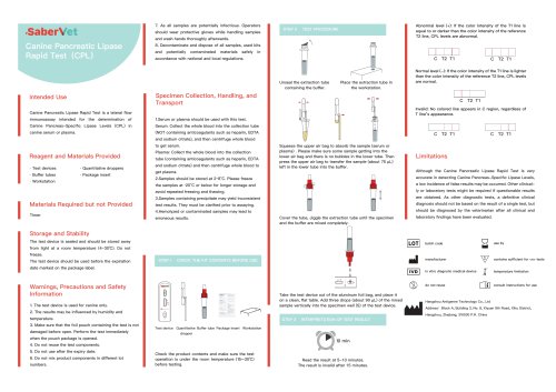
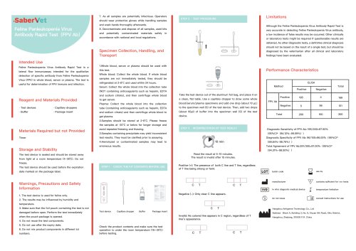
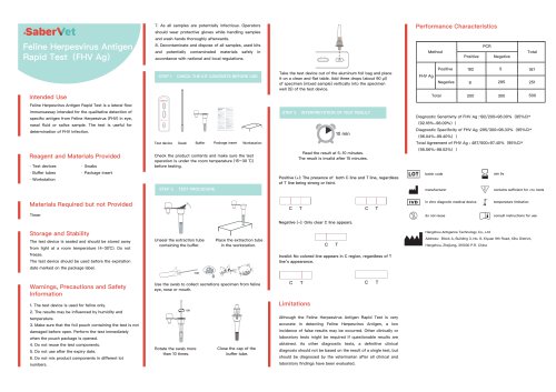
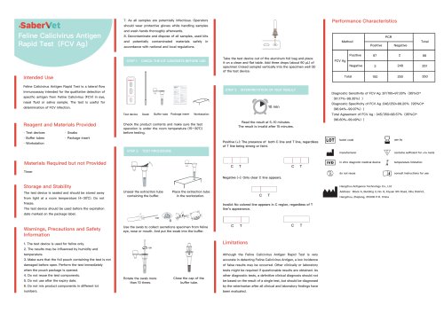
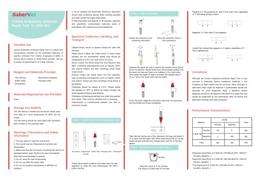
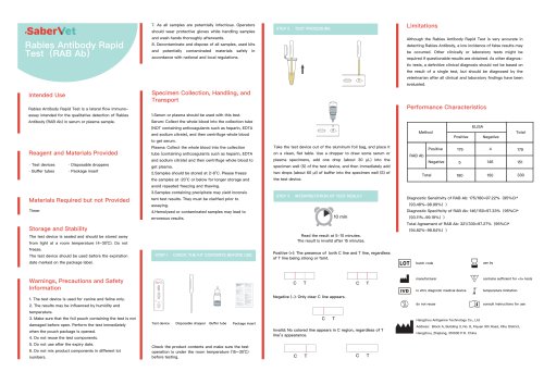
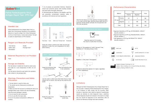
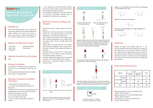
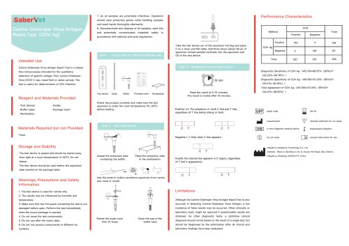
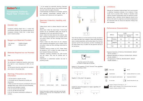
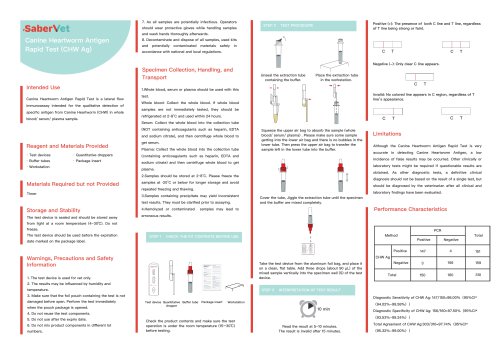
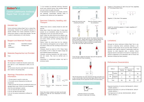
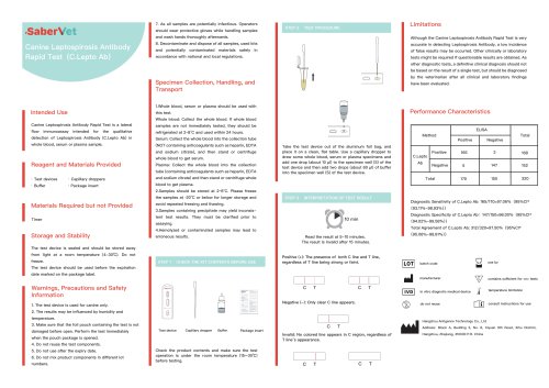
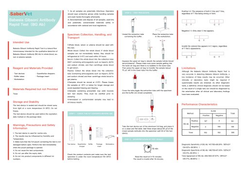
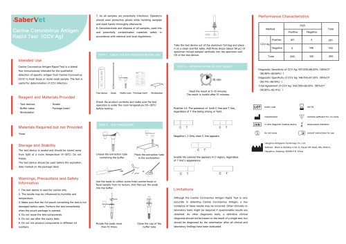
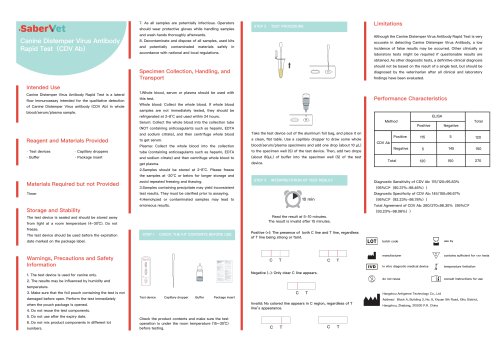
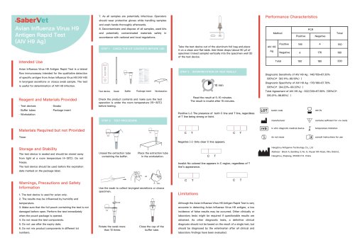
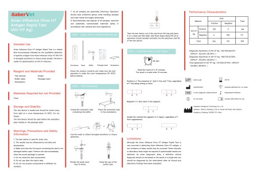
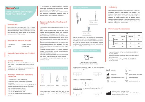
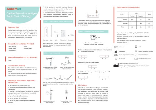
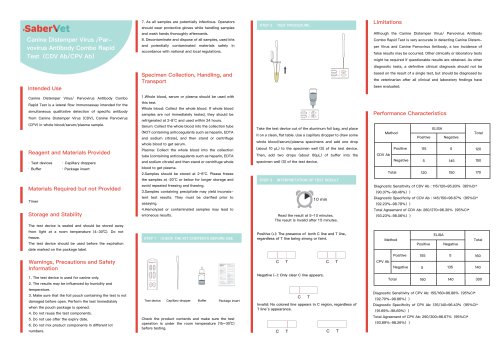
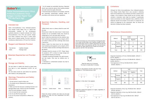
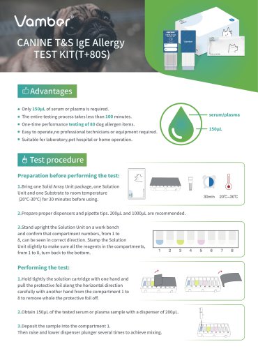
-1-INTENDED USE CANINE T&S IgE TEST KIT is designed to determine the levels of Total IgE (T IgE) and Specific IgE (S IgE) against different allergens in canine serum or plasma. -2-DESIGN AND PRINCIPLE For one sample testing, one Solid Array Unit, one Solution Unit and one Substrate should be used together. The Solid Array Unit, which contains an array composed of location markers, anti-canine IgE antibody, and allergens on a membrane and a protective cap, is packaged in one aluminum foil bag with a desiccant. The Solution Unit contains all the necessary reagents for forming enzyme linked complex of antibody-antigen reaction that are deposited separately in the different compartments of a plastic cartridge and sealed with a protective aluminum foil. The Substrate is deposited in a small substrate bottle. Briefly, pull open the Solution Unit and deposit the serum sample in the compartment 1 of the Solution Unit and mix well. After tearing aluminum foil bag, take the Solid Array Unit out and pull off the protective cap. Immobilized location markers, anticanine IgE antibody, and allergenic substances can be observed as pink spot array on the membrane in the window of the Solid Array Unit. Then insert the Solid Array Unit into the compartment 1 and have it absorb the solution in the compartment 1 for a few minutes. After the absorption, the pink dye will disappear from the membrane in the window, which indicates successful specific antibody-antigen reaction finished. -3- Then the Solid Array Unit will be transferred to the remaining compartments at timed intervals step by step. The bound canine IgE antibodies on the spot array will be labeled with enzyme in the compartment 3, which contains anti-canine IgE-enzyme conjugate. For a satisfactory result, wash steps are introduced. In the compartment 2, the unbound canine IgE antibodies and other substances in the serum sample will be removed. In compartment 4,5and 6,the unbound or excess enzyme conjugate will be adequately removed. At the end, pipette substrate in the substrate bottle, and slowly drop the substrate on the membrane at the window center to develop purple-blue spots if there were enzyme bound there. To confirm the validation of the performance, purple-blue color of the location markers on the membrane should be visible above a certain level after finishing a successful testing process. The location markers will be always visible on the membrane in the window of the Solid Array Unit after successful testing. By placing the transparent Locator on the window of the Solid Array Unit in correct position, the Total IgE and Specific IgE spots can be located. By comparing the visible spots with the Color Scale provided, the signal strength can -4-be obtained and the levels representing the clinical interpretation can be recorded by hand in the Result Card provided according to the INTERPRETING TEST RESULTSTEST PROCEDUREPreparation before performing the test: 1. Bring one Solid Array Unit, one Solution Unit and one Substrate to room temperature (20*C-30*C) for 30 minutes before using. 2. Prepare a dispenser and two pipette tips proper for 200pL and lOOOpL. 3. Stand upright the Solution Unit on a work bench and confirm that compartment numbers, from 1 to 8, can be seen in correct direction. Stamp the Solution Unit slightly to make sure the solutions in the compartments, -5-from 1 to 6, turn back to the bottom.Performing the test: 1. Hold tightly the solution cartridge with one hand and pull the protective foil along the horizontal direction carefully with another hand from the compartment 1 to 8 to remove whole the protective foil off. 2. Obtain 200pL of the tested serum or plasma sample with a proper dispenser set with a pipette tip. EDTA or heparin anticoagulant tubes are recommended for plasma sample collection. -6- 3. Deposit the sample into the compartment 1. Then raise and lower dispenser plunger several times to achieve mixing 4. Tear open the aluminum foil bag, take the solid-state array unit out, and then remove the protective cap. 5. Insert the Solid Array Unit into the compartment 1 for 20 minutes. 6. Pick up the Solid Array Unit and insert it into the compartment 2 for 10 minutes. 7. Pick up the Solid Array Unit and insert it into the compartment 3 for 20 minutes.
Open the catalog to page 1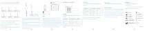
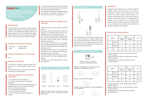
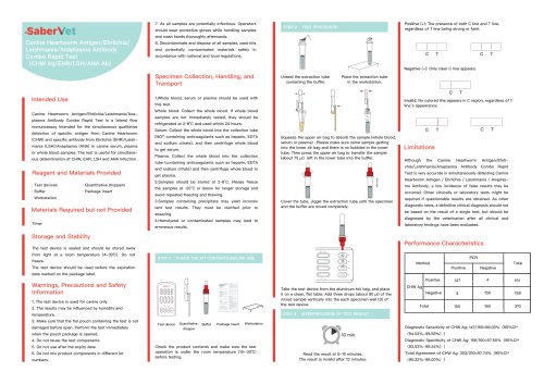
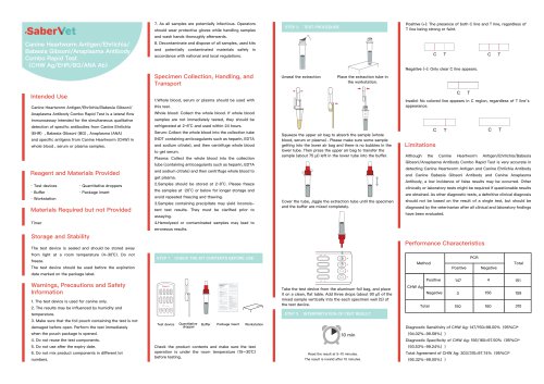
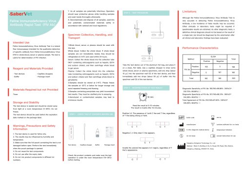
8. Pick up the Solid Array Unit and insert it into the compartment 4 for 10 minutes. 9. Pick up the Solid Array Unit and insert it into the compartment 5 for 10 minutes. INTERPRETING TEST RESULTS Specific IgE Comparing with provided Color Scale, there are four conditions illustrated as the following table. Notes: Total IgE Comparing with provided Color Scale, there are three conditions illustrated as the following table. 10. Pick up the Solid Array Unit and insert it into the compartment 6 for 10 minutes. 11. Pick up the Solid Array Unit and lay it flat on a work bench. 12. Pipette the Substrate...
Open the catalog to page 2All Hangzhou Antigenne Technology Co. Ltd catalogs and technical brochures
 BVDV Ag
1 Page
 BVDV Ag
1 Page
 BRU Ab
1 Page
 FPV FCoV GIA FCV Ag
2 Pages
 FPV FCoV FHV FCV Ag
2 Pages
 CDV CPV CCV GIA Ag
2 Pages
 CDV CAV-II CIV CPIV Ag
2 Pages
 FPV FIPV GIA Ag
2 Pages
 FPV FIPV F.GIA Ag
2 Pages
 FPV FHV FCV Ab
2 Pages
 FPV FCoV FIPV Ag
2 Pages
 FIV Ab FeLV Ag FCoV Ab
2 Pages
 FeLV Ag FIV TOXO Ab
2 Pages
 CHW Ag EHR ANA Ab
2 Pages
 CDV CPV ICH Ab
2 Pages
 FPV GIA Ag
1 Page
 CDV CPV Ag
1 Page
 CDV CPIV Ag
1 Page
 CDV CAV Ag
1 Page
 C.EHR C.ANA Ab
1 Page
 FPV FCoV Ag
1 Page
 ICH Ab
1 Page
 HP Ag
HP Ag1 Page
 GIA Ag
1 Page
 FPL
FPL1 Page
 FHV Ab
1 Page
 FCV Ab
1 Page
 FCoV Ab
1 Page
 F.IgE
F.IgE1 Page
 F-BT A+B
1 Page
 CRV Ag
1 Page
 CAV Ag
1 Page
 C.IgE
C.IgE1 Page
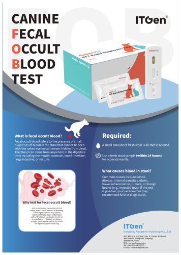
 C.FOB
C.FOB1 Page
 BC Ab
BC Ab1 Page
 FIPV Ag
1 Page
 FeLV Ag
1 Page
 FCoV Ag
1 Page
 F.GIA Ag
1 Page
 RAB Ag
1 Page
 CPV Ab
1 Page
 CIV Ag
1 Page
 CAV-II Ag
1 Page
 CAV-I Ag
1 Page
 SE Ag
SE Ag1 Page
 BRU Ab
1 Page
 FPV/FCoV Ag
1 Page
 CDV CPV CCV GIA Ag
2 Pages
 ITGen catalog
28 Pages
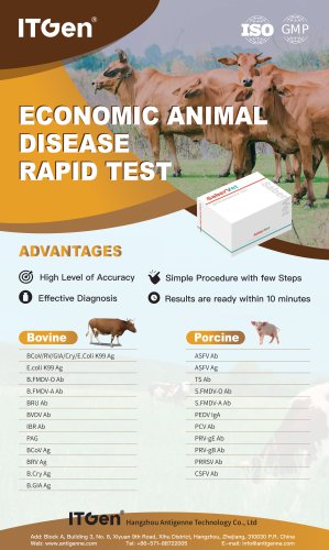 Livestock catalog
1 Page
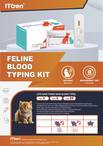 FELINE BLOOD TYPING KIT
1 Page
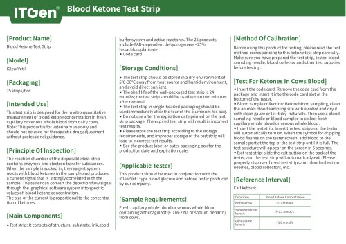
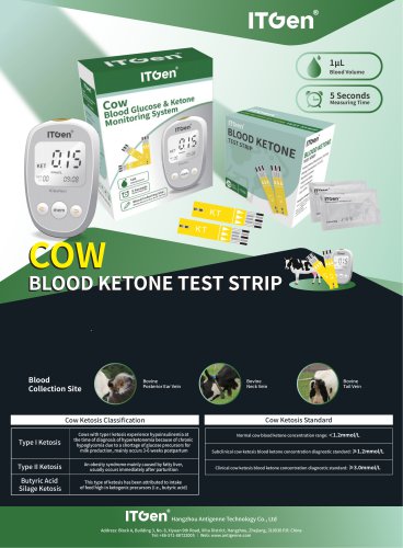 COW BLOOD KETONE TEST STRIP
2 Pages


- Rapid chromatographic immunoassay test
- Blood rapid screening test
- Immunoassay rapid diagnostic test
- Cassette rapid test
- Wound care
- Rapid serum test
- Rapid plasma test
- Rapid virus test
- Rapid whole blood test
- Rapid infectious disease test
- Rapid respiratory disease test
- Urine rapid diagnostic test
- Rapid bacteria test
- Strip rapid screening test
- Bandage
- Fecal rapid test
- Suture thread
- Elastic wound dressing
- Veterinary rapid test
- Rapid nasal test