- Catalogs
- Hermes Medical Solutions, Inc
- NM Processing
NM Processing
1 /13Pages

NM Processing
1 /13Pages
Catalog excerpts
Cardiology Gastroenterology Theranostics & therapy Artificial Intelligence Hepatology Reporting Tools
Open the catalog to page 1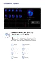
NUCLEAR MEDICINE PROCESSING I Gallbladder ejection fraction analysis pre and post fatty meal Comprehensive Nuclear Medicine Processing at your fingertips ermia offers the most comprehensive Nuclear Medicine processing toolkit on the market. Fully validated applications, for all clinical specialties and all associated image data types, follow the same intuitive steps to facilitate easy adoption within your clinical workflow for:
Open the catalog to page 2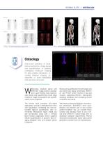
HERMIA SUITE // OSTEOLOGY Tc – 111In Dual Isotope Planar Image Fusion Tc – HDP SUV-SPECT OSEM vs. AMAP-R Advanced Quantitative Reconstruction Osteology End-to-end workflow of multibed reconstruction, image review and quantification in patients undergoing molecular imaging for bone related indications, including infection imaging, assessment of bone metastasis and sacroiliac joint pain. I Sacro-Illiac Joint Analysis. Profile and ROI of Sacro-Illiac Joint hole-body, localized planar and SPECT/CT image review is facilitated by user-friendly, fully customizable viewers and supported by a wide range...
Open the catalog to page 3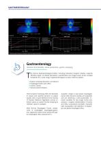
I Salivary gland analysis indications with dual phase acquisition Intestine functionality, saliva production, gastric emptying he Hermia Gastroenterological toolkit, including interactive linogram display, supports effortless region of interest delineation, quantification and image review across multiple timepoints for a wide range of gastroenterological nuclear medicine studies. Gastric emptying (Dynamic and planar) Esophageal transit and reflux Colonic Transit Salivary Gland Analysis Hermia Gastric Emptying offers the flexibility to assess and quantify dynamic or planar studies with in-built reference...
Open the catalog to page 4
HERMIA SUITE // GASTROENTEROLOGY he Hermia Colonic Transit toolkit facilitates fast, regional quantification of patient and fecal imaging and, through geometric center of activity determination, supports effective clinical diagnosis of gut or colonic motility disorders. In-built EANM normal reference values and the option to include custom normal values facilitate easy comparison and diagnostic assessment. With Hermia, manual quantification and spreadsheets can be a thing of the past! Hermia SeHCAT effortlessly and automatically determines background corrected, geometric mean patient counts and...
Open the catalog to page 5
I Future remnant liver function analysis (FRLF) Hepatology Liver function, surgical planning, gallbladder ejection fraction epatobiliary analysis workflow including motion correction algorithms cinematic display, curve plots and customizable results page. These results are readily available through automation. The analysis provides crucial information on the entire biliary system by tracing the production and flow of bile from the formative phase in the liver, and its passage through the biliary system into the small intestine. The application uses a dynamic acquisition of the liver and biliary...
Open the catalog to page 6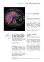
HERMIA SUITE // FUTURE REMNANT LIVER FUNCTION QUICK AND EASY PROCESSING Obtain quantitative results for post-surgical future remnant liver function in minutes I Visualization of the liver remnant volume ENSURE THE BEST OUTCOME FOR YOUR PATIENTS UNDERGOING MAJOR LIVER RESECTION Hermia software analysis allows you to obtain quantitative results to assess post-surgical future remnant liver function in minutes – regardless of the camera vendor. VENDOR NEUTRAL Camera flexibility with standardized workflows Future Remnant Liver Function analysis forms part of the Hermia Nuclear Medicine processing suite,...
Open the catalog to page 7
NUCLEAR MEDICINE PROCESSING // NEPHROLOGY I DMSA Analysis with long-short axis determination and normal database comparison Nephrology Hermia supports DMSA SPECT reconstruction and advanced quantification for all your Nuclear Medicine nephrological investigational needs, tailored to your custom acquisition protocols and personalized to your reporting requirements. ermia offers the flexibility to process all patient cohorts, exhibiting all urological anatomical variations including duplex kidneys and transplant organs, within one user-friendly adaptable platform. The Hermia DMSA toolkit facilitates...
Open the catalog to page 8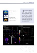
HERMIA SUITE // NEPHROLOGY Renal Curve™ functional images in Hermia a) Uptake Phase Summed Frames b) Patlak Functional Image (slope per pixel) c) Time to Max (Tmax) I Renogram plus post-micturition static analysis with Patlak fit validation ermia seamlessly integrates with Renal Curve™ offering extended renography and post micturition analysis. Improved deconvolution algorithms within Renal Curve™ result in clearer plateaus on retention function graphs and reconvolution curves support quality control of deconvolution analysis. Signal to noise time activity curve smoothing and pseudo-planar functional...
Open the catalog to page 9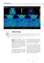
I Subtraction techniques facilitate efficient localization of parathyroid pathology on planar, dynamic or SPECT +/- CT imaging Endocrinology Localization of parathyroid adenoma and Thyroid function assessment. From the origin of nuclear medicine, technologists and physicians have been working on assessing some of the most complex thyroid and parathyroid conditions uantitative assessment of thyroid function in Hermia Thyroid Analysis is possible for acquisition techniques including full/empty syringe imaging, reference capsule and pre-determined efficiency factor. Semi-automated lobar and background...
Open the catalog to page 10
HERMIA SUITE // CARDIOLOGY I Myocardium Gated Analysis (MUGA/FUGA) Cardiology From reconstruction to advanced quantification, Hermia is providing bestin-class product line for cardiology, including third-party software with Invia Corridor4DM and Cedars-Sinai Cardiac Suite. The reconstruction in Hermia improves image quality while reducing dose and acquisition time, minimizing likelihood of patient movement. ermia also features Automatic myocardium detection on SPECT study with the assistance of Artificial intelligence (AI). The algorithm detects the myocardium from anterior and left anterior oblique...
Open the catalog to page 11All Hermes Medical Solutions, Inc catalogs and technical brochures
 Product brochure
36 Pages
 SUV SPECT Reconstruction
4 Pages
 Organ Dosimetry
4 Pages
 Multimodality Viewer
5 Pages
 Pneumology LLQ
3 Pages
 Pneumology VQ
3 Pages
 Neurology
4 Pages
 Voxel Dosimetry
4 Pages
Archived catalogs
 Thyroid PET-CT-MRI
1 Page


- Analysis software
- Visualization software
- Radiology software
- Control software
- Reporting software
- Diagnostic software
- Planning software
- Automated software
- Acquisition software
- Treatment software
- Traceability software
- Data management software
- Surgery software
- Cardiology software
- CT software
- Evaluation software
- 3D viewing software
- Validation software
- Anatomy software
- Quality control software