- Catalogs
- Hermes Medical Solutions, Inc
- Pulmonary Lobe segmentation prior to surgery
Pulmonary Lobe segmentation prior to surgery
1 /1Page

Pulmonary Lobe segmentation prior to surgery
1 /1Page
Catalog excerpts
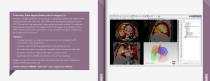
Pulmonary Lobe segmentation prior to surgery (1) Example of image registration of CT perfusion angiography (CTPA) with SPECT-CT VQ study (ventilation and perfusion). The CTPA can be automatically aligned with the PET-CT(s) and the lung segmented using a guided automatic method. The individual lung lobes can be defined in 3D based on the CTPA images, then overlaid onto the SPECT study. Accurate 3D quantification of the individual lobes gives confidence to the surgeon prior to excision. ■ Increased precision by using true lobar anatomy when compared to 2D compartment model approaches ■ Automatic right and left lung segmentation with trachea removal ■ Quick lobe delineation by advanced interactive tools with minimal user input ■ 3D surface rendering of segmented lobes for quality control ■ Export lobe VOIs to DICOM and statistics to Excel™ spreadsheets Images courtesy of Professor Richard Underwood, Royal Brompton Hospital and Harefield NHS Trust, London, UK Software Module: HERMES™ Hybrid 3D™ Lobar Segmentation Module Piqht Lung Left lung NM AC Lung Vent.urt.on SP£CT w*h SC /£! : 21 M-„ NM AC Lung Perfusion SPECT CT with SC 21 ^ Load Daiwt
Open the catalog to page 1All Hermes Medical Solutions, Inc catalogs and technical brochures
 Product brochure
36 Pages
 SUV SPECT Reconstruction
4 Pages
 Organ Dosimetry
4 Pages
 Multimodality Viewer
5 Pages
 Pneumology LLQ
3 Pages
 Pneumology VQ
3 Pages
 Neurology
4 Pages
 NM Processing
13 Pages
 Voxel Dosimetry
4 Pages
Archived catalogs
 Thyroid PET-CT-MRI
1 Page


- Analysis software
- Visualization software
- Radiology software
- Control software
- Reporting software
- Diagnostic software
- Planning software
- Automated software
- Acquisition software
- Treatment software
- Traceability software
- Data management software
- Surgery software
- Cardiology software
- CT software
- Evaluation software
- 3D viewing software
- Validation software
- Anatomy software
- Quality control software