- Catalogs
- Hermes Medical Solutions, Inc
- Thyroid PET-CT-MRI
Thyroid PET-CT-MRI
1 /1Page

Thyroid PET-CT-MRI
1 /1Page
Catalog excerpts
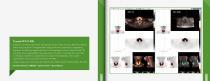
Thyroid PET-CT-MRI Example of a 55 year old female with thyroid cancer. A head and neck PET-CT combined with a head and neck Tl-weighted MRI. Using the Hermes automatic co-registration algorithm, the MRI was aligned with the CT and displayed in fusion mode with the PET. Automatic lesion segmentation was performed on the PET to calculate the mean SUV in the lesion, max SUV and peak SUV. As a result of the registration, the volume segmentation can also be seen overlaid on the MRI. Images courtesy of Dr. Annick Van den Abbeele, Dana Farber Cancer Institute, Boston, MA, USA Software Module: HERMES™ Hybrid Viewer™ Base Module
Open the catalog to page 1All Hermes Medical Solutions, Inc catalogs and technical brochures
 Product brochure
36 Pages
 SUV SPECT Reconstruction
4 Pages
 Organ Dosimetry
4 Pages
 Multimodality Viewer
5 Pages
 Pneumology LLQ
3 Pages
 Pneumology VQ
3 Pages
 Neurology
4 Pages
 NM Processing
13 Pages
 Voxel Dosimetry
4 Pages
Archived catalogs


Related Searches
- Analysis software
- Visualization software
- Radiology software
- Control software
- Reporting software
- Diagnostic software
- Planning software
- Automated software
- Treatment software
- Acquisition software
- Traceability software
- Data management software
- Surgery software
- Cardiology software
- CT software
- Evaluation software
- 3D viewing software
- Validation software
- Anatomy software
- Quality control software
*Prices are pre-tax. They exclude delivery charges and customs duties and do not include additional charges for installation or activation options. Prices are indicative only and may vary by country, with changes to the cost of raw materials and exchange rates.