
- Catalogs
- Hitachi High-Tech Europe GmbH
- SU8700 | Ultra high resolution FE SEM


- Company
- Products
- Catalogs
- News & Trends
- Exhibitions
SU8700 | Ultra high resolution FE SEM
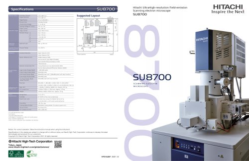
SU8700 | Ultra high resolution FE SEM
Catalog excerpts
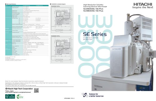
Schottky Emitter Accelerating Voltage Probe Current Stage Control Suggested Layout Step-down Transformer (Option) Specimen Chamber Specimen Size Standard Detectors Operation table Upper Detector (UD) Lower Detector (LD) Variable Pressure Pressure Range (VP) mode (*2) Movable Range Specimen Stage Secondary Electron Image resolution Electron Optics Hitachi UltraHigh-resolution Field-emission Scanning electron microscope Customer supplied item Middle Detector (MD) Semiconductor Type BSED (PD-BSED) Ultra Variable-Pressure Detector (UVD) STEM Detector Energy Dispersive X-ray Spectrometer (EDS) Electron Backscattered Diffraction Detector (EBSD) Image Display Mode Large Screen Display Mode Single Image Display Mode Dual Image Display Mode 800×600 pixels and 1,280×960 pixels with dual monitors Quad Image Display Mode Six Image Display Mode w/dual monitors 640×480 pixels with dual monitors Pixel Size SU8700 SCANNING ELECTRON MICROSCOPE Utility Requirements Main Unit Power(main unit) 4 kVA(crimp contact for M6)AC100 V±10 %, or AC200-240 V ±10 % with autotransformer Cooling Water (Chiller) Dedicated Cooling Water Circulation system (*5) Vacuum Pump with deceleration mode Option Mountable Detectores Weight of standard unit; does not include options. Customer-supplied item In case of connection from installation site facilities. Notice: For correct operation, follow the instruction manual when using the instrument. Specifications in this catalog are subject to change with or without notice, as Hitachi High-Tech Corporation continues to develop the latest technologies and products for our customers. Copyright (C) Hitachi High-Tech Corporation 2
Open the catalog to page 1
SU8700 Modern Solutions for a Modern World The SU8700 brings in a new era of Ultrahigh-resolution Schottky field emission scanning electron microscopes to the long-standing Hitachi EM line-up. This revolutionary FE-SEM platform incorporates multifaceted imaging, high-probe current, automation, efficient workflows for users of all experience levels, and more. Enhanced User-Experience with Advanced Automation . Automated alignments increase efficiency and throughput. . Automated data acquisition recipes allow for greater precision as well as repeatability. . High-precision piezo stage* improves...
Open the catalog to page 2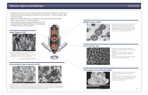
Electron Optics and Detectors The SU8700 allows for both versatile imaging and demanding analytical capabilities in a single system. . Schottky field emitter provides probe current from a few pA to 200 nA, in order to address a large variety of specimens. . Novel electron optics enable very low voltage imaging while still maintaining high image quality – even as low as 0.1 kV without stage bias. . Optimized detection system and chamber design allow short working distance EDS analysis (WD=6 mm) for both imaging and analysis under the same parameters. Backscatter electron (BSE) imaging is a powerful...
Open the catalog to page 3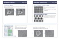
Automated Workflows Application - Semiconductor - Automated Optics Alignment Voltage Contrast Images of 7 nm process SRAM SEM operation requires optimization of various parameters when conditions, specimens, or analyses change. The SU8700 features an automated alignment function to assist in this procedure. From beam alignment to stigmator alignment, each alignment optimization can be done automatically. Voltage contrast in a SEM is a very powerful tool for semiconductor device evaluation. As the device structure changes by layer, SEM conditions such as accelerating voltage and contrast must...
Open the catalog to page 4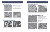
Application - Material Science - Tempered Martensite in Steel Application - Life Science - Ultrastructure of Arabidopsis thaliana Detailed evaluation of metallic alloys is very important to many industries. The left two images demonstrate the strong capabilities of the SU8700 for such investigations. The precipitates along grain boundaries are clearly visible when acquiring secondary electron images using the UD. Grain size and deformation are easily distinguishable in left-bottom image by acquiring BSE channeling contrast. In addition, crystal defects in some grains can be clearly observed in...
Open the catalog to page 5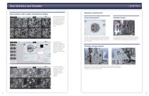
User Interface and Chamber Operation assisting GUI Dual monitors with 6-signal simultaneous display Multi-Channel image can be displayed on the screen. 2, 4 or 6 signals including the chamber scope(*) or SEM MAP image can be displayed simultaneously on a single monitor. (top) The dual-monitor configuration supports enhanced productivity plus expanded workspace and allows the operation panel to be customized with submenus positioned anywhere on either screen. (center) In multi-signal display mode, a saved image can be loaded to a vacant channel and further processed for real-time comparison to...
Open the catalog to page 6All Hitachi High-Tech Europe GmbH catalogs and technical brochures
 SU8700
12 Pages
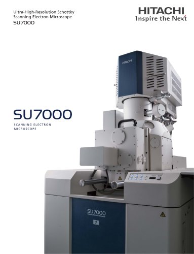 SU7000
8 Pages
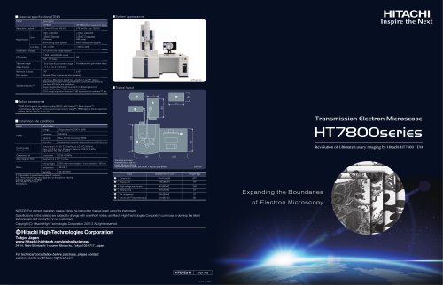 HT7800 Series | TEM
2 Pages

Archived catalogs
- Analysis software
- HITACHI laboratory microscope
- Desktop microscope
- Sample preparation system
- Automatic sample processor
- Control software
- Laboratory software
- Laboratory sample preparation system
- Benchtop sample preparation system
- Acquisition software
- Biology microscope
- Spectrophotometer
- HITACHI research microscope
- Image analysis software
- Research software
- HITACHI high-resolution microscope
- Medical microscope
- Compact microscope
- Inspection microscope