HOCT-1/1F
1 /12Pages
HOCT-1/1F
1 /12Pages
Catalog excerpts

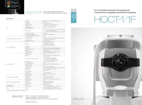
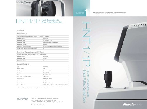
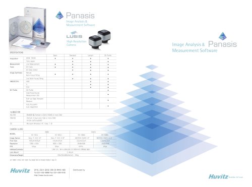
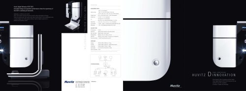
All-in-One Optical Coherence Tomography with Fundus Camera, Angiography, Biometry and Topography Specification Principle Spectral domain OCT, Fundus digital photography Light source Scan speed 20 um (Lateral), 7 um (z-axis) at index 1.36 Scan Range Display resolution Minimum pupil diameter Scan patterns Macular : Macular Line, Macular Cross, Macular Radial, Macular3D, Macular Raster, Angio (Option) Disk : Disc Circle, Disc Radial, Disc 3D, Disc Raster, Angio (Option) Optical power at cornea Acquisition time of 3D image Depth Accuracy (measuring 1 mm glass) Angiography Range OCT Angiography – Option (HOCT-1, HOCT-1F) Superficial, Deep, Outer, Choroicapilary, Retina,Custom, Enface, Thickness map, Depth coded map Angiography Analysis Non-mydriatic fundus camera 60 line pair/mm or more (center) 40 line pair/mm or more (middle) 25 line pair/mm or more (periphery) Built-in 12M pixel, Color or Built-in 20M pixel, Color Minimum pupil diameter 4.0 mm (Normal mode), 3.3 mm (Small pupil mode) Flash light White light, 10 levels Pixel pitch at fundus 3.69 um (20M pixel Color)4.63 um (12M pixel Color) Capture mode Single, Stereo, Widefield Panorama Working distance 12.1 inch, 1280 x 800 pixel, Touch panel color LCD Dioptric compensation forpatient’eye s -33D~+33D total -13D~+13D with no compensation lens +7D~+33D with plus compensation lens -33D~-7D with minus compensation lens Fixation target LCD (internal), White LED (external) Fundus illumination light Common specification Horizontal movement 70 mm (back and forth), 100 mm (left and right) Vertical movement Chinrest movement Auto tracking 30 mm (up and down), 10 mm (right and left), 10 mm (back and forth) Power supply External port Dimensions / Mass Anterior segment adapter (optional) Working distance Scan range 6 ~ 9 mm (width), 2.3 mm (depth) ACA line, Anterior Radial Software Analysis Corneal Layers, Thickness Map, Thickness, Angle Working distance Wide Anterior segment adapter (optional) Scan pattern Scan range Scan pattern ACA line, Anterior Radial, Full Software Analysis Dimension, Angle Biometry (optional) Topography (optional) Supported Maps Axial map, Tangential map, Keratoconus Screening Web-Based, Multi users can be accessible Progression analysis, Comparison analysis, 3D Analysis * Specification and design are subject to change without notice. HUVITZ Co., Ltd. 38, Burim-ro 170beon-gil, Dongan-gu, Anyang-si, Gyeonggi-do, 14055, Republic of Korea Tel: +82-31-442-8868 Fax : +82-31-477-8617 http ://www.huvitz.com W2ARAA-19-00001 Rev.2 All-in-One Optical Coherence Tomography with Fundus Camera, Angiography, Biometry and Topography
Open the catalog to page 1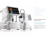
3D OCT FUNDUS CAMERA ANGIOGRAPHY BIOMETRY TOPOGRAPHY See more, Do more, Save more Huvitz 5-in-1 OCT 3D OCT with Fundus Camera and Angiography HOCT is now even more advanced with the addition of Biometry and Topography. Not only Anterior and Posterior disease diagnosis, but also gathering the necessary data for an Ophthalmologist’ s cataract surgery. Because the HOCT acquires all the necessary information in one instrument, it becomes efficient and convenient for you and your patients.
Open the catalog to page 2
Segmentation of Seven Retinal Layers High-Speed & High-Quality Incredible speed of 68,000 A-scan/sec. : More Realistic and Clearer image in high resolution B-scan and 3D Image of Macular Provides High-speed Scan, High-quality Image by using Huvitz's outstanding optical technology and innovative image software. Shows extensive information, such as 3D structure of Retina, Macula's thickness and separation in a vivid image. High Resolution Image - min. 60 lines/mm of central Fundus Creates 3 um OCT Digital Resolution medical images, allows more precise Retina observation and useful follow-up examinations....
Open the catalog to page 3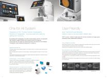
User Friendly Integration of OCT, Fundus Camera, Angiography, Biometry & Topography : more accurate and useful by adding all 5 functions. Auto Tracking & Auto Shooting : makes it easy to use and obtain reliable data HOCT is smart.- Obtains reliable data with minimum deviation of image quality according to user's measurement proficiency. By combining OCT Angiography, Full Color Fundus Camera, and PC, it can generate high resolution images providing multi-purpose functions for diagnosis. It saves both time and space by performing frontal view (Enface) of eye diseases, Tomography, cross-compare...
Open the catalog to page 4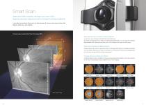
Smart Scan Start and finish instantly through only one-click : Speedy process reduces errors in forward looking of patients It provides convenience & accuracy by offering easy & various scanning functions with Macula, Optic Disc, and Anterior. Concept image visualized Smart Scan Technology (SST). Wide Area Scan (12 mm x 9 mm) for efficient diagnosis 12 mm A quick scan covers Macula and Optic Disk areas extensively. By scanning around Optic Disc or Macula for patient's pathological status, you can check the Thickness Maps between RNFL (Retinal Nerve Fiber Layer), GCL (Ganglion Cell Layer) and...
Open the catalog to page 5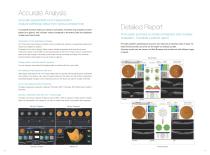
Accurate Analysis Accurate segmentation and measurement : Analyze pathology status from various perspectives A complete analysis helps you observe symptoms, illnesses and progress of each patient at a glance. Key indicator values compared to Normative Data are displayed in table and chart format. Detailed Report From quick summary to simple comparison and complex evaluation : Complete a perfect report Progression to track pathological changes OCT scan and fundus image of a patient can be compared at a glance to sequential measurement results from baseline to present. Progression from past to...
Open the catalog to page 6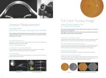
Full Color Fundus Image Anterior Measurement One Single System : Start and finish in one place, making patient more comfortable Anterior Segment Module allows measurement and analysis of cornea thickness, angle and 3D image. It helps users work more efficiently by acquiring both anterior and posterior in one place. Insight of Posterior Segment of Eye : for Comprehensive diagnosis Color Retinal Images optimized with high-resolution and contrast are very useful in analysis and clinical diagnosis. Best images are provided by Low intensity of flash, fast capture speed, quiet operation, small pupil...
Open the catalog to page 7All Huvitz catalogs and technical brochures


Archived catalogs
 HDC-900N
2 Pages
 HRK-9000A
4 Pages
 Imaging Solution
2 Pages
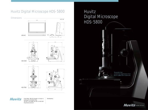 HDS-5800 Series
8 Pages
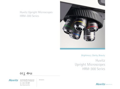 HRM-300 Series
10 Pages
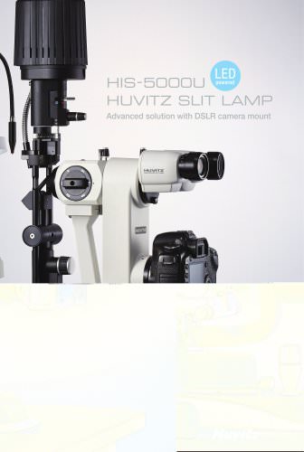 HIS-5000U
2 Pages
 HBK-7000
2 Pages
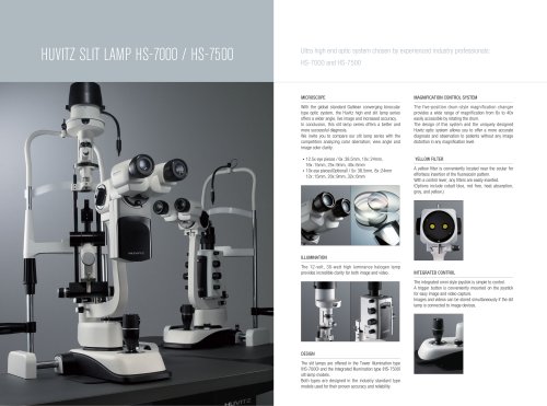 HS-7000
1 Page
 HS-5000
2 Pages
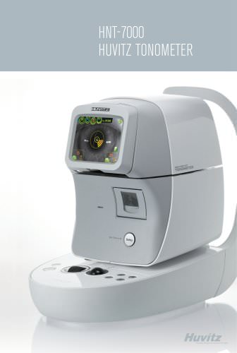 HNT-7000
2 Pages
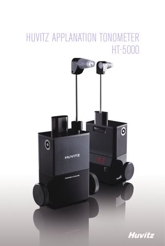 HT-5000
2 Pages
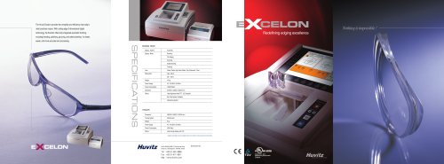 EXCELON
2 Pages
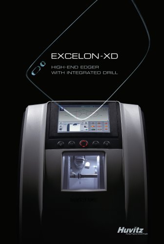 EXCELON XD/XQ
8 Pages
 CAB-4000
2 Pages
 CLM-3100P
2 Pages
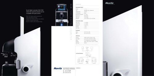 HLM - 7000
2 Pages
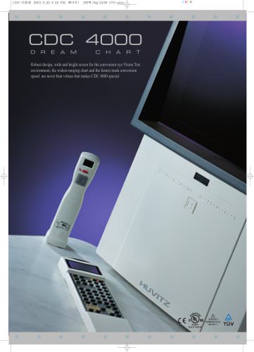 CDC-4000
2 Pages
 HCD-7000
2 Pages
 CCP-3100
2 Pages
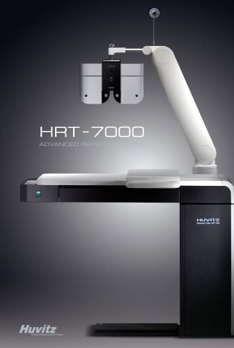 HRT-7000
2 Pages
 HDR-7000
2 Pages
 HRK-7000A
2 Pages
 HRK-8000A
2 Pages

- Analysis software
- Tablet computer software
- Tablet PC software
- Diagnostic software
- Dental software
- Fixed ophthalmic examination
- Design software
- 3D printer
- Acquisition software
- Treatment software
- Dental milling machine
- Data management software
- Measurement software
- CAD/CAM milling machine
- 3D scanner
- 3D dental scanner
- Dental 3D printer
- Import software
- Server software
- Workstation with chair