- Catalogs
- Idexx Laboratories
- Computed Radiography in Perspective
- Products
- Catalogs
- News & Trends
- Exhibitions
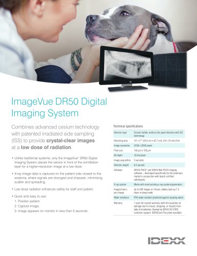
Computed Radiography in Perspective

Computed Radiography in Perspective
The document provides a historical context of radiography, starting with the discovery of X-rays by Wilhelm Conrad Roentgen in 1895, which revolutionized medical diagnostics by enabling noninvasive visualization of bones.
X-ray Generator
Radiography systems, including film-screen radiography (FSR), computed radiography (CR), and direct digital radiography (DDR), rely on an X-ray generator. The generator's output is managed by adjusting milliampere (mA), exposure time, and kilovolt peak (kVp). High-frequency generators are preferred for their consistent output and reduced motion blur.
Conventional Film Screen Radiography (FSR)
FSR captures images on film after X-rays pass through a patient. Image density is affected by mA and exposure time, while contrast is influenced by kVp. The ALARA principle is emphasized to minimize radiation exposure. Intensifying screens and grids enhance image quality and reduce scatter radiation.
Common Causes of Image Problems
Image quality issues in FSR can result from technical errors like improper film density, motion blur, and exposure problems, as well as contaminated chemicals and environmental factors.
Computed Radiography (CR)
CR creates digital images using an imaging plate (IP) and reader, involving image acquisition, processing, and display. CR is increasingly popular in veterinary practices due to affordability and ease of use. CR equipment includes an IP, reader, and computer with software for image manipulation and storage.
CR Equipment and Image Quality
CR systems require high-resolution monitors for accurate diagnosis. While CR reduces film and development errors, it can still introduce artifacts. CR is more forgiving of exposure errors than FSR, allowing greater latitude in image correction.
Transition from FSR to CR
Transitioning from FSR to CR can reduce retakes and radiation exposure but requires careful calibration and technique chart development to optimize exposure levels. Studies show CR can reduce radiation doses without compromising diagnostic information.
Specifications and Procedures
CR systems enhance visualization of different tissue densities, useful for comparing images of the lungs and heart. Picture Archiving and Communication Systems (PACS) software improves diagnostic capabilities but requires careful handling to avoid artifacts.
Artifacts and Solutions
Common artifacts during the transition from FSR to CR include thin white lines and general fogging. Solutions involve replacing cracked imaging plates, applying lead foil to cassettes, and ensuring proper loading of imaging plates.
Advantages of Computed Radiography
CR offers benefits over FSR, such as reduced costs for chemicals and film, elimination of darkroom space, and fewer technical errors. It supports mobile veterinary practices by enabling on-site diagnosis and treatment, reducing follow-up visits.
Direct Digital Radiography (DDR)
DDR is similar to CR but acquires images directly without an imaging plate and reader. While DDR quality is comparable to CR, CR remains preferred in some settings due to its established use and reliability.
Recommendations
To minimize artifacts, limit the number of individuals involved in image post-processing and rely on default software settings. The transition to digital radiography is seen as inevitable and beneficial for veterinary practices.
Conclusion
The transition to CR and DDR is a significant investment but offers long-term benefits in cost, efficiency, and diagnostic capabilities. Training and proper equipment handling are crucial to maximizing these benefits.
Acknowledgments
The author acknowledges contributions from various professionals and references several studies and guidelines to support the findings and recommendations.
Catalog excerpts
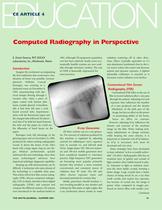
Computed Radiography in Perspective E. David Stearns, RVT, IDEXX Laboratories, lnc.,Westbrook, Maine Imagine the excitement accompanying the first realization that noninvasive visu- alization of bones was possible. German physicist Wilhelm Conrad darkened room on November 8, \ 1895, experimenting with elee- \ trical charges flowing through a \ vacuum tube, when a piece of \ paper coated with barium plat- \ inum cyanide glowed. Coincident- \ ally, it had been left near a card- \ board covered tube. Experiment- \ ation with the fluorescent paper and \ the charged tube followed. He discov- \ ered that if he held his hand between \ the tube and the paper, he could see V the silhouette of hand bones on the Roentgen took full advantage of the glowing paper and on December 22,1895, produced the oldest existing radiographic record. It shows the bones of his wife's hand with a large signet ring on one fin- ger. Medical professionals quickly embraced this new technology. Since then many technological advances have improved radiologic diagnostic capability. Keeping up with advancements can be difficult. A technician who stays abreast of the technology is a valuable clinic asset. This article will review film-screen radiog- raphy (FSR), discuss computed radiogra- phy (CR) in depth, introduce direct digital compare the different systems. CR systems were introduced to the medical market in 1981. Although CR equipment acquisition cost has been relatively steady, more eco- nomically feasible systems are now avail- able through veterinary vendors. The cost most practices. _ X-ray Generator All three systems use an x-ray genera- tor. The amount of radiation produced by this machine is regulated by adjusting controls for milliampere (mA), exposure time in seconds (s), and kilovolt peak ary and 100-mA mobile generators have been considered standard in veterinary practice. High frequency (HF) generators are becoming more popular primarily because they produce a more constant, concentrated, and consistent source of radiation than SP units. The HF unit offers shorter exposure times and decreased potential for motion blur. Ideally, generators should produce x- rays traveling parallel to one another and striking the film plane at right angles. But x-ray production always results in some radiation scattering off in other direc- tions. Filters (typically equivalent to 2.5 mm aluminum) positioned close to the x- ray source reduce this scatter and decrease patient and operator exposure. A lighted adjustable collimator is essential as it decreases scatter radiation even further. Conventional Film Screen i film to record radiation after x-rays pass \ through the patient. Adjusting mA and I exposure time influences the number ft of x-rays produced and the density ■ (blackness) of the dark part of the ■ image, but has no affect on the power M or penetrating ability of the beam, ^ hence no affect on contrast. However, adjusting kVp influences the density and contrast of the resultant image on the film. When making tech- nique adjustments to change contrast, while keeping the same density, it is important to maintain mAs-kVp balance. decreased and vice versa. Many strategies have been developed to keep radiation "as low as readily achiev- able" (ALARA). Radiographic film has an emulsion layer of gelatin and crystals of light-sensitive silver halide bound to plas- tic. Exposure to light or radiation sensi- tizes the silver halide crystals, forming a latent image. Large crystals have a better chance of being struck by an x-ray than small crystals and require less exposure, but because of crystal size, the image is grainy when compared to images pro- duced on slower films with smaller crys- THE NAVTA JOURNAL • SUMMER 2004
Open the catalog to page 1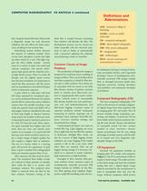
COMPUTED RADIOGRAPHY CE ARTICLE 4 continued tals. Using the fastest film that will provide a diagnostic image not only decreases radiation, but also allows for faster expo- sures, resulting in less motion blur. Intensifying screens further decrease the amount of radiation needed. These screens have phosphor crystals that light up when struck by x-rays. This light trig- gers the silver halide crystals. Screens come in a variety of types (rare earth and calcium tungstate) and speeds (ultra- speed, high-speed and the relatively slow- er high-detail screens). There is a trade-off though, and the...
Open the catalog to page 2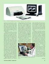
Figure I Computed Radiography equipment includes an imaging plate, reader; and computer archiving and communication systems (PACS) are provided with some veterinary systems. PACS not only manage image processing and display, but also control data storage, retrieval and transfer. PACS are most useful when they interface with existing practice management software, allowing attachment of the image to the patient record. The IP, which looks like a conventional intensifying screen, has a layer of crystals that can store x-ray energy. This plate is placed in a cassette, similar to the conven- tional...
Open the catalog to page 3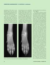
COMPUTED RADIOGRAPHY CE ARTICLE 4 continued underexposure of film results in a non- diagnostic image in most cases, whereas the same level of CR underexposure is eas- ily managed by imaging software. CR lati- tude for overexposure is even greater. With CR, errors made setting the machine have to be substantially out of range to require retake. A corrected over- exposed image will have more detail than a normally exposed image. Correction of underexposed images boosts contrast, but causes decreased signal-to-noise ratio, resulting in decreased detail. Radiologists making a diagnosis from an image...
Open the catalog to page 4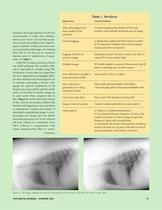
situations, but to get radiation ALARA it is recommended to make dose measure- ments as part of an x-ray machine accept- ance test and, if possible, to use a high fre- quency machine. Artifacts are more com- mon during the initial stages of switching operator error or malfunction of equip- Using PACS image processing software can clarify pathology that would be diffi- cult or impossible to visualize using FSR Production of more than one image from the same digital data can highlight differ- ent tissue densities and aid in diagnosis. As an example, processing a thoracic radi- ograph for optimal...
Open the catalog to page 5All Idexx Laboratories catalogs and technical brochures
Archived catalogs
 IDEXX VetStat
86 Pages
 Pseudalert
8 Pages
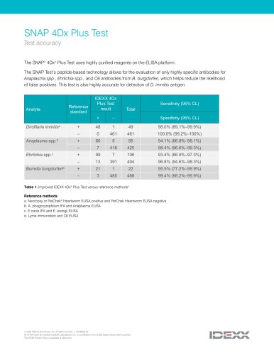
 IDEXX SNAP 4Dx Plus
2 Pages
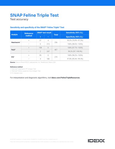
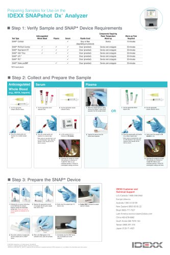
 SNAPshot® DSR
2 Pages
 ELISA Tips
1 Page
 Digital Imaging Brochure
10 Pages


- Detection kit
- Blood detection kit
- Serum detection kit
- Immunoassay detection kit
- Blood rapid screening test
- IDEXX rapid immunoassay test
- Infectious disease detection kit
- IDEXX cassette rapid test
- Rapid serum test
- Rapid plasma test
- Rapid virus test
- Rapid whole blood test
- IDEXX rapid infectious disease test
- Tablet computer software
- Tablet PC software
- Clinical chemistry analyzer
- ELISA detection kit
- Automated biochemistry analyzer
- Benchtop biochemistry analyzer
- IDEXX rapid bacteria test