- Catalogs
- IMAGELEVEL
- Morita 3D Accuitomo 170
Morita 3D Accuitomo 170

Morita 3D Accuitomo 170
The document introduces a new imaging technology with an 80 μm voxel resolution, providing high-quality images with low x-ray radiation exposure. This technology aims to deliver detailed imaging while minimizing patient dose.
The system offers four imaging modes to meet various diagnostic needs:
- High Resolution Mode: Provides the highest spatial resolution, ideal for observing delicate bone structures.
- High Fidelity Mode: Offers high data density for clearer images, suitable for zoom reconstructions.
- High Speed Mode: Reduces motion artifacts, beneficial for patients who have difficulty remaining still.
- Standard Mode: Suitable for both limited and broad fields of view.
The system provides nine field of view sizes, ranging from 170 mm to 40 mm in diameter, to minimize x-ray dosage. Five resolution levels are available, allowing selection of voxel sizes from 80 μm to 250 μm, depending on diagnostic needs.
Zoom reconstruction enables detailed examination of critical areas using voxel sizes as small as 80 μm, enhancing image resolution.
The compact design requires minimal space, with recommended room dimensions provided. The system is DICOM compatible and includes viewing software for manipulating 3D-CT image data.
The system operates by rotating an arm 360° around the exposure region, emitting a cone-shaped x-ray beam. The resulting projections are converted into digital signals, processed, and reconstructed into 3D CT images.
The x-ray dosage for a standard 18-second exposure is significantly lower than that of conventional CT scans, enhancing patient safety.
The isotropic cubic voxel ensures high-resolution images with minimal artifacts, maintaining image quality across all dimensions.
The FPD enhances image quality and reduces x-ray dosage. It is unaffected by magnetic fields and provides high sensitivity and resolution.
The i-Dixel software offers extensive image processing capabilities, including volume rendering, real-time re-slice, and curved MPR. It supports DICOM standards and facilitates image sharing across clinic networks.
This advanced imaging technology provides high-quality, detailed 3D-CT images with reduced radiation exposure, making it a valuable tool for diagnostic imaging.
The document provides technical specifications and operational procedures for the 3D Accuitomo 170, a high-resolution imaging device developed by J. Morita Mfg. Corp. It includes details on software compatibility, positioning systems, imaging capabilities, and technical specifications.
The 3D Accuitomo 170 can be integrated into external clinic networks using i-Dixel Viewer Software, One Data Viewer, and One Volume Viewer, allowing 3D-CT images to be viewed on PCs even without the device present.
The Scout Positioning System is highlighted for its simplicity and accuracy, utilizing a triple beam system for precise targeting. It allows for easy targeting of the region of interest with two-direction scout imaging, minimizing x-ray dosage by accurately determining the minimal region of interest.
The device offers various imaging area options with different diameters and heights, allowing for detailed examination of specific regions such as the paranasal sinuses and temporal bone. High-resolution zoom reconstruction is available with voxel sizes as small as 80 μm for detailed observation.
The 3D Accuitomo 170 operates with a tube voltage range of 60-90 kV and a tube current of 1-10 mA. It supports multiple exposure modes, including Standard, Hi-Fi, Hi-Res, and Hi-Speed, with varying exposure times. The field of view options range from ø 170 x 120 mm to ø 40 x 40 mm, with voxel sizes from 80 μm to 250 μm.
The main unit dimensions are 1,620 mm x 1,250 mm x 2,080 mm, and it weighs approximately 400 kg. The control box dimensions are 96 mm x 40 mm x 115 mm. The device requires a power supply of 100/110/120VAC or 220/230/240VAC with a maximum power consumption of 2.0 kVA.
The device is developed and manufactured by J. Morita Mfg. Corp., located in Kyoto, Japan. Technical assistance is provided by NubiC, and the device is a result of collaborative development with Nihon University.
Catalog excerpts

Super Resolution: 80 μm Voxel Low Patient Effective Dose High Quality Images with Low X-Ray Radiation Four Imaging Modes A mode to serve every purpose. High Resolution Mode and High Fidelity Mode can be used for even higher quality images. High Speed Mode reduces motion artifacts. Use Standard Mode for both limited and broad fields of view. Nine Sizes for Field of View (FOV) Choose from nine sizes for the FOV with diameters ranging from170 mm to 40 mm to minimize x-ray dosage. Five Resolution Levels Select the voxel size, 80 μm, 125 μm, 160 μm, 200 μm, or 250 μm, that best suits your diagnostic...
Open the catalog to page 4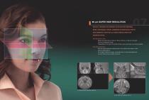
80 μm Super High Resolution Four Imaging Modes Select a region of interest such as the temporal High Resolution Mode and High Fidelity Mode can be used bone, paranasal sinUs, jawbone or individual teeth for even higher quality images. High speed mode reduces motion artifacts. and observe it with 80 μm voxel resolution for Use Standard Mode for both limited and broad fields of view. greater detail. High Resolution Mode (Hi-Res) Five Resolution Levels This is the highest resolution. Exposures are made at one-fourth the size of Select voxel size, 80 μm, 125 μm, 160 μm, 200 μm, or 250 μm, that best...
Open the catalog to page 5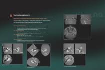
80 μm Super High Resolution Four Imaging Modes Select a region of interest such as the temporal High Resolution Mode and High Fidelity Mode can be used bone, paranasal sinUs, jawbone or individual teeth for even higher quality images. High speed mode reduces motion artifacts. and observe it with 80 μm voxel resolution for Use Standard Mode for both limited and broad fields of view. greater detail. High Resolution Mode (Hi-Res) Five Resolution Levels This is the highest resolution. Exposures are made at one-fourth the size of Select voxel size, 80 μm, 125 μm, 160 μm, 200 μm, or 250 μm, that best...
Open the catalog to page 6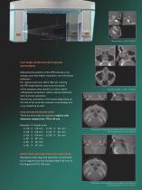
Cone Beam CT Radiography The arm rotates 360° around the center of the exposure region Flat Panel Detector (FPD) Position in 18 seconds (Standard Mode) as the x-ray head emits a cone-shaped beam. The multiple projections created during the arm’s rotation are converted to a digital signal by the Adjusting the position of the FPD reduces x-ray flat panel detector and transmitted to the computer. After dosage, provides higher resolution, and minimizes any necessary supplemental or corrective processing, the digital information is converted into a three dimensional CT For regions such as ø 140 X...
Open the catalog to page 7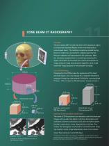
Cone Beam CT Radiography The arm rotates 360° around the center of the exposure region Flat Panel Detector (FPD) Position in 18 seconds (Standard Mode) as the x-ray head emits a cone-shaped beam. The multiple projections created during the arm’s rotation are converted to a digital signal by the Adjusting the position of the FPD reduces x-ray flat panel detector and transmitted to the computer. After dosage, provides higher resolution, and minimizes any necessary supplemental or corrective processing, the digital information is converted into a three dimensional CT For regions such as ø 140 X...
Open the catalog to page 8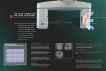
High Quality 3D-CT Images with Low X-Ray Radiation Using a high-sensitivity, high-resolution Digital Signal a-Si (amorphous silicon) array flat panel detector, high quality and extremely detailed images of the many regions of the head and neck such as the temporal bone, paranasal sinuses, eye sockets, mandible, and cranial base can be obtained for a wide range of multi-purpose diagnostic scanning. Flat Panel Detector (FPD) necessity of making corrections for distortion before reconstructing image quality and a reduction in x-ray dosage. The flatness of the detector minimizes distortion. This...
Open the catalog to page 9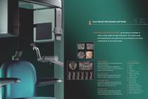
i-Dixel Image Processing Software i-Dixel image processing software can be used as a database to archive a wide variety of image information. Its multiple image processing functions can easily access and manipulate many types of information for 2D and 3D images. Volume Rendering Volume rendering of CT data produces three dimensional images. Select the area of interest and adjust the controls for the histogram to create a detailed image of very fine stractures. Histogram Edge Enhancement Real Time Re-Slice Slices and volume rendered images can be linked and easily manipulated in real time. Distance...
Open the catalog to page 10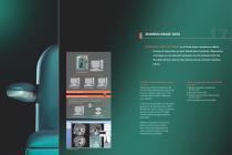
SHARING IMAGE DATA INSTALLING i-Dixel SOFTWARE on all intra-clinic computers enables sharing of image data on each linked client computer. Observation of images on non-network computers can be achieved with the One Data Viewer, and the One Volume Viewer without installing i-Dixel. One Data Viewer & One Volume Viewer Software i-Dixel software Out of network computer In external clinic networks without 3D Accuitomo 170, 3 D-CT images can be viewed on a PC with both of the following methods: 2. Storage service class computer does not have i-Dixel soft- Intra-clinic network 1. Modality worklist management...
Open the catalog to page 11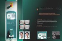
Simple, Accurate Positioning The Scout Positioning system is easy and accurate. Use the Triple Beam positioning system for even greater Precision. Two-Direction Scout The region of interest can be easily targeted by making images from two directions. Then you can simply click on the images to specify the center of the region of interest. This information is transmitted to the x-ray unit, and the chair automatically moves into position. The Scout exposure (80 kV and 2.0 mA) will increase the total x-ray dosage of a Standard Mode CT exposure (90 kV and 5.0 mA) by about 2%. Easy High Precision The...
Open the catalog to page 12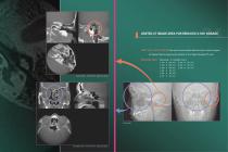
Limited CT Image Area for Reduced X-Ray Dosage Limit the x-ray dosage Use scout to accurately determine the minimal region of interest before exposing the patient to the highter dosage CT scan Imaging Area : Diameter X Height (mm) ø 170 X 120 mm Scout Image Paranasal sinuses φ170 X 120 mm. Voxel size: 250 μm Temporal Bone. φ60 X 60 mm. Voxel size: 125 μm
Open the catalog to page 13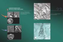
Zoom Reconstruction with 80 μm Voxel Resolution Select a region of interest such as the temporal bone or paranasal sinus and zoom in with 80 μm voxel resolution for a Volume rendering produces a detailed 3D more detailed observation. view of internal structures. Auditory ossicular chain Auditory ossicular chain zoom reconstruction Ethmoid sinus zoom reconstruction Lamina Cribrosa
Open the catalog to page 14All IMAGELEVEL catalogs and technical brochures
 Morita IC5-HD
6 Pages
 The new Mediadent MDX3
1 Page
 Morita 3D R100
9 Pages
 VistaScan Perio
6 Pages
 VistaScan Combi
8 Pages
 Veraview iX Intraoral X-ray
2 Pages
 VistaCam camera systems
6 Pages
 Mediadent V6
3 Pages
 D.F.O.
1 Page
 Mediadent
2 Pages


