- Catalogs
- IMAGELEVEL
- Morita 3D R100
Morita 3D R100

Morita 3D R100
The Veraviewepocs 3D R100 and F40 models represent a new frontier in X-ray diagnostics, offering innovative 3D Reuleaux Full Arch Fields of View (FOVs) that enhance dental imaging capabilities. These models are designed for a variety of dental applications, including implant planning, with a focus on reducing dosage and improving image clarity.
Specifications and Features
- 3D Reuleaux Full Arch FOV: The R100 model features a unique convex triangle shape for full arch imaging, reducing dosage by excluding unnecessary areas. It offers six FOV options ranging from 40 x 40 mm to 100 x 80 mm.
- High Resolution and Dose Reduction: Both models provide high-resolution images with a voxel size of 0.125 mm. The Dose Reduction Mode optimizes X-ray intensity, reducing exposure by up to 40% compared to standard modes.
- Positioning and Imaging: The system includes easy 3D positioning with panoramic images, bi-directional scout, and five laser beams for precise alignment. The C-arm automatically adjusts to the optimal position for imaging.
- Software and Image Processing: The i-Dixel 2.0 software supports advanced implant planning and is compatible with third-party software. Features include cMPR image processing, mandibular canal tracing, and an implant library for realistic presentations.
Clinical Applications
- Endodontics: The 3D R100 model provides detailed images for diagnosing and planning treatment for complex cases, such as apical lesions and sinus membrane perforations.
- Implantology: Offers comprehensive imaging for implant planning and post-operative observation, ensuring high-quality bone assessment and implant health.
- Oral Surgery: The system aids in diagnosing impacted teeth and associated complications, providing clear views of bone loss and sinus damage.
Technical Specifications
- Dimensions and Power: The main unit measures W 1,020 x D 1,300 x H 2,355 mm, with a power consumption of 2.3 kVA.
- X-ray Generator: Tube voltage ranges from 60-90 kV, and tube current from 1-10 mA, depending on the exposure mode.
- Imaging Areas: The R100 model offers imaging areas from Ø 40 mm x H 40 mm to Ø 100 mm x H 80 mm, while the F40 model provides Ø 40 mm x H 40 mm and Ø 40 mm x H 80 mm.
Conclusion
The Veraviewepocs 3D R100 and F40 models are advanced diagnostic tools that offer flexibility, high resolution, and reduced dosage for a wide range of dental applications. Their innovative design and software capabilities make them ideal for modern dental practices.
Catalog excerpts
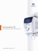
Veraviewepocs 3D F40 and R100 with innovative 3D Reuleaux FOV Thinking ahead. Focused on life.
Open the catalog to page 1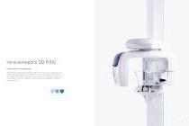
Veraviewepocs 3D R100 A New Frontier in X-ray Diagnostics Veraviewepocs 3D R100 has changed the shape of 3D. This unit's groundbreaking and patent pending 3D Reuleaux Full Arch elds of view (FOVs) provide a unique shape for full arch imaging. With 6 eld of view options and Morita's world renowned image quality, Veraviewepocs 3D R100 is suitable for a wide variety of dental applications including implant planning.
Open the catalog to page 2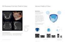
3D Reuleaux Full Arch Field of View Various Fields of View Exposure Areas for Multiple Diagnostics The Veraviewepocs 3D R100 model offers a total of 6 exposure areas from 40 x 40 mm up to 100 x 80 mm for various diagnostic needs. The new full arch scan captures the maxilla and/or the mandible with the equivalent of 100 mm in diameter and two height options of 50 or 80 mm. Its full arch capability, reduced dosage and exceptional clarity are ideal features for implant planning and oral surgery. This unit also offers small and medium eld of view sizes suitable for endodontics, periodontics, as well...
Open the catalog to page 3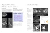
High Resolution Images With Dose Reduction Feature Dose Reduction Feature Through advanced engineering, a Dose Reduction Mode optimizes the intensity of the X-rays which lowers exposure for easily penetrated tissues. Dosage is reduced to a mere 40 % of the standard mode.* By maximizing efciency, soft tissue, such as the maxillary sinus membrane and skin, appear sharper than ever before with fewer artifacts.** Resolution & Clarity Veraviewepocs offers high resolution images of 125 µm voxel. It provides clear images of the periodontal pocket, the periodontal ligament, and the alveolar bone. It...
Open the catalog to page 4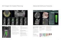
3D Images for Implant Planning Planning Process Successful placement of implants starts with the very critical and detailed planning process dentification of structures such as the sinus cavity, inferior alveolar nerve and clear views of the bone for implant planning with full arch imaging, industry leading clarity, and i-Dixel 2.0 software offers advanced implant planning features, plus compatibility with popular third party cMPR Image Processing Create cross sectional images of the dental arch Mandibular Canal Tracing Highlight the mandibular cana for easier viewing, measuring determining its...
Open the catalog to page 5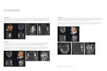
Clinical Cases Implantology The patient was seen for a routine follow up visit following implant placement of tooth #16. The implant had been placed 9 years earlier. The coronal, sagittal, and axial views all conrm good quality bone around the implant and an absence of any pathology related to the treatment. The ne detail of the bone, especially in the coronal view, allows the clinician and the patient to feel condent about the current health of the implant. Oral Surgery The patient presented with pain in the maxillary left region. A cone beam CT image was taken with the 3D R100 and it was revealed...
Open the catalog to page 6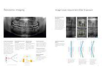
Panoramic Imaging Image Layer Adjustment After Exposure Panoramic Image Layer Adjustment The image layer for panoramic images can be adjusted after the exposure has been made to improve clarity and sharpness. The focus can be improved for points of varying depth as well as the surface. Select any point in the image for focus enhancement and then use the mouse wheel to make the adjustment. After Image Layer Adjustment AF Automatic Positioning This function makes patient positioning nearly effortless. A light beam sensor automatically positions the unit without requiring the patient to move. The...
Open the catalog to page 7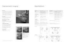
Cephalometric Imaging High Speed The Veraviewepocs system offers high speed performance requiring only 4.9 seconds for a cephalometric scan. The speed helps ensure high quality images each and every time. For pediatric patients, the reduced scan time is especially helpful as repeat images due to patient movement are virtually eliminated. Trade name: Model: Type: Panoramic image Exposure time: Input voltage: Power consumption: Standard Panoramic (standard, orthogonal and shadow reduction projections) Magnication: 1.3 X throughout and 1.6 X throughout Dimensions Main unit: With Cephalometric: Pedodontic...
Open the catalog to page 8
Diagnostic and Imaging Equipment Treatment Units Handpieces and Instruments Endodontic Systems Laser Equipment Laboratory Devices Developed and Manufactured by: J. Morita Mfg. Corporation 680 Higashihama Minami-cho, Fushimi-ku, Kyoto, 612-8533 Japan T +81 (0)75. 611 2141, F +81 (0)75. 622 4595 www.jmorita-mfg.com Morita Global Website www.morita.com J. Morita Corporation 33-18, 3-Chome, Tarumi-cho Suita City, Osaka, 564-8650 Japan T +81 (0)6. 6380 1521, F +81 (0)6. 6380 0585 J. Morita USA, Inc. 9 Mason lrvine, CA 92618, USA T +1 (0)949. 581 9600, F +1 (0)949. 465 1095 J. Morita Europe GmbH Justus-von-Liebig-Str....
Open the catalog to page 9All IMAGELEVEL catalogs and technical brochures
 Morita IC5-HD
6 Pages
 The new Mediadent MDX3
1 Page
 Morita 3D Accuitomo 170
17 Pages
 VistaScan Perio
6 Pages
 VistaScan Combi
8 Pages
 Veraview iX Intraoral X-ray
2 Pages
 VistaCam camera systems
6 Pages
 Mediadent V6
3 Pages
 D.F.O.
1 Page
 Mediadent
2 Pages


