- Catalogs
- INFINITT North America
- INFINITT_XELIS
INFINITT_XELIS

INFINITT_XELIS
- Offers 2D, 3D, and 4D tools for Coronary CT Angiography and Ventricle Analysis.
- Enhanced CT Colonography for fast polyp detection.
- Supports multi-modality study fusion and 3D image visualization.
- Provides reliable color maps with quantitative results.
- Flexible licensing options: full package or specialized modules.
- Seamless integration with INFINITT PACS.
- Regular software upgrades and concurrent user access.
- Server: Intel Quad-core CPU x 2, 16 GB RAM, 10 TB HDD, Windows Server 2008 R2.
- Client: Intel Pentium II 300MHz, 512MB RAM, 2GB HDD, Windows XP SP3.
- Xelis Cardiac: Tools for Coronary CT Angiography with features like automatic rib cage removal and vessel tracking.
- Xelis Colon: CT colonography solution with real-time navigation and polyp detection.
- Xelis Lung: Pulmonary nodule detection with over 80% detection rate for nodules.
- Xelis Fusion: Multi-modality image fusion with SUV analysis for PET-CT images.
- Xelis Perfusion: Blood flow imaging application for brain perfusion analysis.
Catalog excerpts
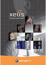
3D Diagnosis Support System
Open the catalog to page 1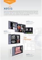
This advanced visualization software is designed for better data Loading and reconstruction of large-volume datasets, while the Web-based technology enables access from any Internet-connected or wireless PC. The name Xelis comes from 'xel' denoting image element, as in pixel (2D) and voxel (3D), and 'IS' for information systems. Volume rendering and other post-processing techniques for enhanced visualization, treatment planning, and workflow for Infinitt's Web-based PACS. Xel?S Cardiac Easyand powerful 2D,3D,and 4D tools Left/Right Ventricle Analysis Enables advanced 3D visualization and analysis...
Open the catalog to page 2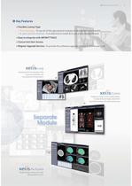
3D Diagnosis Support System Key Features Flexible License Type Full package : To use all of the specialized modules to be economically feasible A specialized module : It is selective to install & to use a specialized module Easy to integrate with INFINITT PACS Concurrent User Access Regular Upgrade Service : To provide the software upgrade service 2~3 times per year Essential tool for the evaluation of the lung and lung nodules, and the comparison of serial exams Enables you to fuse multi-modality studies, visualize 3D fused images and perform quantitative analysis with speed and accuracy Reliable...
Open the catalog to page 3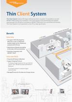
Thin Client System Thin Client System realizes 3D images without any time or any place. It is possible to use with INFINITT PACS without any installation. Thin Client System can be downloaded and updated automatically if the 3D option is activated on INFINITT PACS and It can use all Xelis functionality. Thin Client System is not required for a client PC as high specication. Benet Xelis Only To support 2D & 3D altogether. Wherever a user is, Diagnostic Quality image can be created and analyzed with 3D tools. Rapid Volume Rendering loading & All kinds of 3D functionalities Seamless integration...
Open the catalog to page 4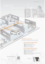
3D Diagnosis Support System DICOM Image Data Captured Image & Job Save Data Server Hardware Specification Client Hardware Specification • CPU: Intel Pentium II 300MHz higher • Display: 1280 x 1024 LCD Display resolution is required
Open the catalog to page 5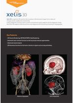
Xelis 3D is a general 3D solution that visualizes 3 dimensional images from a data set of large volume rapidly and conveniently. It has a diagnostic function and it is useful to nd lesions and to apply for all the diagnostic elds. Also Xelis 3D supports effective and accurate diagnosis with the post-processing for Angiography, Key Features - 3D Volume Rendering, MIP/MinIP/MPR, Slab Rendering - Automatic bone removal function as well as precise manual segmentation - Advanced vessel analysis - 3D Volumetry function for the tumor volume or organs such as lung and kidney Abdomen Angiography Brain...
Open the catalog to page 6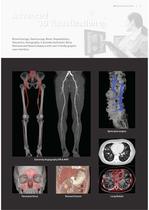
3D Diagnosis Support System Bronchoscopy, Gastroscopy, Bone, Hepatobiliary, Volumetry, Venography. It provides Automatic Bone Removal and Vessel Analysis with user friendly graphic user interface. Spine post surgery Paranasal Sinus Stomach Cancer Lung Nodule
Open the catalog to page 7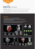
Xelis Cardiac provides easy and powerful 2D, 3D, and 4D display and analysis tools for evaluation of Coronary CT Angiography. It is easy and powerful 2D, 3D, and 4D display tool for evaluation of coronary CT angiography. Automatic removal of rib cage, auricle, and ventricle help to improve productivity with only one-click vessel tracking. Key Features - Automatic Cardiac Axis view - Chamber view (HLA,VLA,SA) - Powerful MPR slab review including 4D Cardiac MPR and VR image to visualize cardiac wall motion and contractility - Automatic blood pool segmentation, detection of LV/RV contours and LV/RV...
Open the catalog to page 8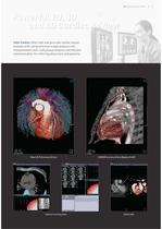
3D Diagnosis Support System Xelis Cardiac offers fast and accurate cardiac vessel analysis with comprehensive image analysis and measurement tools, soft plaque analysis and efcient communication for referring physicians and patients. Heart & Pulmonary Artery Calcium scoring view CABG(Coronary Artery Bypass Graft) Valve view
Open the catalog to page 9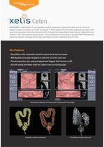
Xelis Colon is a dedicated CT colonography solution designed to improve the detection of colorectal polyps through correlation of 2D and 3D images. It offers greater efciency and diagnostic accuracy with real-time navigation tools and endoluminal y-through techniques (band view) that can display the inner surface of the colon without any distortion. These visualization techniques, combined with workow and reporting features, are expected to help radiologists shorten reading time by 20~30%. Key Features - Easy-Dene-Path : Automatic centerline extraction for each air-bowel - With Bandview one-pass...
Open the catalog to page 10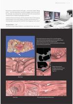
3D Diagnosis Support System “Panoramic endoluminal y-through - commonly called ‘Band View’ - and the seamless overlay of tagged stool during 3D y-through, are substantial advances from the conventional endoluminal or attening display methods. I believe these techniques will be of great help in decreasing interpretation time and making 3D post-processing of CT colonography cases performed with stool tagging much easier” Seong Ho Park | MD University of Ulsan College of Medicine, Asan Medical Center, Seoul, Korea The surgical specimen shows a 3.5-cm ulcerofungating adenocarcinoma (arrowheads) and...
Open the catalog to page 11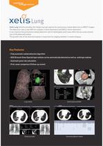
Xelis Lung Solution provides the helpful second-opinion for pulmonary nodule detection on MDCT images. The detection rate is over 80% for nodules (›3mm diameter) and GGOs (›6mm diameter). It can improve the pulmonary nodule detection rate of radiologists and it also offers the accurate volume size of the detected nodule. The growth rate of the detected nodule is important for judging whether it needs a biopsy. Key Features - Fully automatic nodule detection algorithm - GGO (Ground-Glass Opacity) type nodules can be automatically detected as well as solid type nodules - Automatic grow rate calculation...
Open the catalog to page 12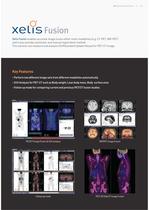
3D Diagnosis Support System Xelis Fusion enables accurate image fusion either multi-modalities (e.g. CT-PET, MR-PET) and it also provides automatic and manual registration method. This solution can measure and analysis SUV(Standard Uptake Values) for PET-CT image. Key Features - Perform two different image sets from different modalities automatically - SUV Analysis for PET-CT such as Body weight, Lean body mass, Body surface area - Follow up mode for comparing current and previous PET/CT fusion studies PET/CT Image Fusion & SUV analysis MR/PET Image Fusion Prior exam Current exam PET/ 3D Slab...
Open the catalog to page 13All INFINITT North America catalogs and technical brochures
 INFINITT_C_PACS_Brochuere
4 Pages
- Analysis software
- Radiology software
- Reporting software
- Laboratory software
- Monitoring software
- Diagnostic software
- Planning software
- Cloud-based software
- Hospital software
- Dental software
- Treatment software
- Data management software
- AI-assisted software
- Measurement software
- Artificial intelligence software
- Surgery software
- Web-based software
- Simulation software
- Sharing software