
- Catalogs
- Innomed Medical
- PI-PACS Chest CAD


PI-PACS Chest CAD

PI-PACS Chest CAD
Catalog excerpts
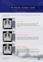
PI-PACS Chest CAD Decision Support System for Nodule Detection on Lung The PI-PACS Chest CAD assists you in finding, quantifying and reporting lesions on lung fields in digital chest X-ray images. Second eye Built in CAD system for Chest X-ray images is available in PI-PACS offering a real time second opinion assisting early detection. Advanced visualisation enables quick and convenient workflow at your workstation. Soft tissue analysing Missed lung cancer might be the consequence of anatomical structures (e.g. collarbone, rib) covering lung nodules on a chest X-Ray. Bone shadow eliminating algorithms make the lung tissue visible. Nodule detection Dens, circular shapes or blurred, infiltrated areas that can be the early sign of tumour or TB are marked. The identified lesions are segmented directing attention to suspicious areas. To find these symptoms as small as possible is crucial in early treatment. [email protected] www.innomed.hu
Open the catalog to page 1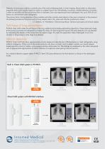
Detection of pulmonary nodules is certainly one of the most challenging tasks in chest imaging. Based solely on attenuation properties even small nodules might be visible on a digital chest X-ray. Nevertheless in practice, reliable detection of nodules is often not possible until they are at least 8mm in diameter. Even very experienced chest radiologists miss up to 30% of such lesions on conventional chest radiographs. The primary factor limiting detection of lung nodules and other similarly sized objects is their poor conspicuity in the presence of overlaying anatomical structures such as lung...
Open the catalog to page 2All Innomed Medical catalogs and technical brochures



Archived catalogs
 ArguSys++
2 Pages
 InnoCare-T12
2 Pages
 HeartScreen 80G-L
2 Pages
 HeartScreen 80 GL-1
2 Pages
 HeartScreen 112 C-1
2 Pages
 Cardio PC/E
2 Pages
 Cardio-Aid 360B_2019
2 Pages
 InnoCare-T17 Plus
2 Pages
 TOP-X 100LC
2 Pages
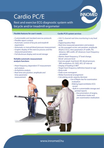 CardioPC/E
2 Pages
 Cardio-Aid 360B_2013
2 Pages
 TOP-X 100NR generator
2 Pages
 Cardio-Aid TM 360-B
2 Pages
 HeartScreen 210
2 Pages
 InnoCare-T12 Plus
2 Pages
 TOP-X 100NR
2 Pages
 CardioAid-1
2 Pages
 HeartScreen 60-IKO
2 Pages
 HeartScreen 60G
2 Pages
 HeartScreen 80 CL
2 Pages
 CardioAid 200B
3 Pages
 InnoCare VET
2 Pages
 HeartScreen 60G VET
2 Pages
 InnoCare-S
2 Pages
 HeartScreen 112Clinic
4 Pages
 HeartScreen 80C-L
2 Pages
 InnoCare-CC
2 Pages


- 1-door wardrobe
- Digital radiography system
- 1-unit wardrobe
- Medical device cabinet
- Digital electrocardiograph
- EKG
- Multipurpose radiography system
- X-ray system
- White cabinet
- Wall-mounted cabinet
- Multipurpose radiography X-ray system
- Multi-parameter monitor
- Training manikin
- External defibrillator
- Multi-parameter O2 monitor
- Pulse oximetry multi-parameter monitor
- Multi-parameter ECG monitor
- Blood pressure multi-parameter monitor
- Compact multi-parameter monitor
- 12-channel electrocardiograph