- Catalogs
- Integra LifeSciences
- DIABETIC SKIN ULCER
DIABETIC SKIN ULCER

DIABETIC SKIN ULCER
- INTEGRA™ Matrix Wound Dressing: A porous matrix made of cross-linked bovine tendon collagen and glycosaminoglycan, designed to support cellular invasion and capillary growth.
- INTEGRA™ Bilayer Matrix Wound Dressing: Similar to the Matrix Wound Dressing but includes a semi-permeable silicone layer for enhanced tear strength and moisture control.
- Indicated for partial and full-thickness wounds, pressure ulcers, venous ulcers, diabetic ulcers, and more.
- Contraindicated for patients with sensitivity to bovine collagen or chondroitin materials and not suitable for third-degree burns.
- Do not resterilize or use if the package is damaged.
- Ensure control of exudate, bleeding, swelling, and infection before application.
- Possible complications include infection, chronic inflammation, allergic reactions, and excessive redness or swelling.
Catalog excerpts
INTEGRA™ MATRIX WOUND DRESSING • INTEGRA™ BILAYER MATRIX WOUND DRESSING SOFT TISSUE SOLUTIONS Collagen Soft T issue Integra™ Extremity Reconstruction
Open the catalog to page 1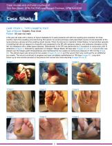
Case studies and pictures courtesy of: Tzvi Bar-David, DPM FACFAS and Robert Fridman, DPM AACFAS Case Study CASE STUDY 1 - TYPE II DIABETIC FOOT Type of Wound: Diabetic Foot Ulcer Patient: 58 year-old male A 58-year old male with a history of Type-II diabetes for 8 years presents with left foot swelling and ulceration for three months. Each time swelling occurred during this period, he would purchase a self-prescribed course of oral Ampicillin at the local pharmacy abroad and the swelling would recede. When he returned to mainland United States, he had another episode of swelling that did not...
Open the catalog to page 2
Case Study CASE STUDY 2 - TYPE II DIABETIC FOOT Type of Wound: Diabetic Foot Ulcer Patient: 61 year-old male A 61 year-old male, with a medical history notable for Type II diabetes, hypertension and hypercholesterolemia, presented with an ulcer on the lateral aspect of the left midfoot. On physical exam, there was an abscess which tracked medially across the entire plantar midfoot (Image #1-2). The patient was admitted emergently for antibiosis, and the wound was debrided and abscess drained in the OR (Image#3). The right lower extremity was subsequently revascularized. The tunneled track was...
Open the catalog to page 3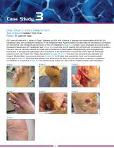
Case Study CASE STUDY 3 - TYPE II DIABETIC FOOT Type of Wound: Diabetic Foot Ulcer Patient: 67 year-old male A 67-year old male with a history of Type II diabetes and HIV, with a history of abscess and osteomyelitis of the left 5th metatarsal head, with subsequent resection of the metatarsal head. Approximately two years later he developed a contracted 4th hammertoe with retrograde plantar flexion of the 4th metatarsal (Image #1). A grade II ulcer developed as a result of the increased pressure sub-4th metatarsal head (Image #2). Local care and off-loading with orthotics did not resolve the problem....
Open the catalog to page 4
Case Study CASE STUDY 4 - TYPE II DIABETIC FOOT Type of Wound: Diabetic Foot Ulcer Patient: 54 year-old male A 54-year-old male with multiple medical problems most notable for diabetes on hemodialysis, hypertension, coronary artery disease, and peripheral vascular disease, presented upon referral with a 3 month history of dry gangrene of the right halux. He recently had an arthrectomy of the right popliteal artery, and subsequently developed the gangrene. An MRI showed evidence of an abscess extending from the area of dry gangrene demarcation to the plantar vault with a swelling and malodor (Image...
Open the catalog to page 5
Case Study CASE STUDY 5 - TYPE II DIABETIC FOOT Type of Wound: Diabetic Foot Ulcer Patient: 80 year-old male An 80-year old male with a medical history notable for Type-II diabetes presented to the emergency room with a right foot ulcer draining plantarly and medially for 1 week. The ulcers had been present for approximately 10 years, and had been only treated conservatively. On physical exam, there was extreme pain on palpation of the right foot with malodor and copious yellow-gray drainage. The patient had previous digital amputations bilaterally. Upon evaluation, the prognosis of the right...
Open the catalog to page 6
A PICC line was placed for long-term IV antibiotics for treatment of osteomyelitis with Vancomycin and Imipenem. Approximately one month later, the wound bed was prepared, and Integra™ Bilayer Matrix was applied as per standard protocol (Images #5-8). IV antibiotics were completed and non-weight bearing status was obtained through the use of a wheelchair. There was adequate wound epithelialization at 113 days after Integra application for the patient to advance to ambulation in custom molded shoes and inserts (Images #9-11). There was complete epithelialization of the wound at 155 days after...
Open the catalog to page 7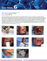
Case Study CASE STUDY 6 - TYPE II DIABETIC FOOT Type of Wound: Diabetic Foot Ulcer Patient: 53 year-old male A 53-year old male with a history of uncontrolled Type II diabetes presented with a one month history of enlarging heel ulcer with purulent drainage after a puncture wound on a metal surface (Image #1). Plain film x-rays showed no break in the calcaneal cortex, however, a Technesium bone scan showed increased uptake in the late phase. The patient had operative debridement of necrotic tissue, and inspection of the calcaneus showed hard bone with no evidence suggestive for osteomyelitis....
Open the catalog to page 8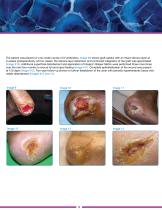
The patient was placed on a six-week course of IV antibiotics. Image #9 shows graft uptake with an intact silicone layer at 2-weeks postoperatively. At four weeks, the silicone layer detached, and continued integration of the graft was appreciated (Image #10). Additional superficial debridement and application of Integra™ Bilayer Matrix were performed three more times over the next four months to ensure full and rapid healing (Image #11). Complete epithelialization of the wound was present at 153 days (Image #12). Two-year follow-up shows no further breakdown of the ulcer with periodic hyperkeratotic...
Open the catalog to page 9
Case Study CASE STUDY 7 - TYPE II DIABETIC FOOT Type of Wound: Diabetic Foot Ulcer Patient: 55 year-old male A 55-year old male with a history of uncontrolled Type-II diabetes, hypertension, end-stage renal disease on hemodialysis, presents with ulceration of the left foot with gas gangrene. He underwent a transmetatarsal amputation which progressed to dehiscence of the entire wound. He was treated with local wound care with topical debridement agents without success. Below-knee amputation was considered, and he was referred for attempt at limb salvage. Wound cultures positive for pseudomonas...
Open the catalog to page 10
The wound showed uptake of the graft with formation of neo-dermis (Images #6 and 7). Two additional applications of Integra™ Bilayer Matrix were applied at 51 and 99 days post-operatively (Image #8) with progression of healing (Image #9). Complete epithelialization of the wound was achieved at 161 days (Image #10 and 11). Image 6
Open the catalog to page 11
Integra™ Matrix Wound Dressing Integra™ Bilayer Matrix Wound Dressing Indications for Use: INTEGRA™ MATRIX WOUND DRESSING Description INTEGRA™ Matrix Wound Dressing is an advanced wound care device comprised of a porous matrix of cross-linked bovine tendon collagen and glycosaminoglycan. The collagen-glycosaminoglycan biodegradable matrix provides a scaffold for cellular invasion and capillary growth. Indications INTEGRA™ Matrix Wound Dressing is indicated for the management of wounds including: partial and full thickness wounds, pressure ulcers, venous ulcers, diabetic ulcers, chronic vascular...
Open the catalog to page 12All Integra LifeSciences catalogs and technical brochures
 capture high-torque surgical
12 Pages
 cadence
4 Pages
 DuraGen® Secure
8 Pages
 transducer
2 Pages
 MAYFIELD®
12 Pages
 SurgiMend® Hernia Brochure
8 Pages
 AmnioExcel® OR Sell Sheet
2 Pages
 PyroCarbon Implants
8 Pages
 Wrist Solutions
6 Pages
 Lower Extremity Reconstruction
28 Pages
 Integra® Lighting Solutions
4 Pages
 Integra® CUSA® Excel+
1 Page
 CUSA® Excel+ System
4 Pages
 DuraGen® matrix
2 Pages
 SurgiMend®
6 Pages
 Integra® Tissue Technologies
8 Pages
 Auragen™ Cortical Electrodes
4 Pages
 DuraGen XS
2 Pages
 Suturable DuraGen™
4 Pages


Archived catalogs
 MAYFIELD® 2
3 Pages
 DuraGen Plus
4 Pages
 Hermetic Plus™
2 Pages
 AccuDrain™
4 Pages
 Oral Surgery
107 Pages
 Catalog - Endodontics
110 Pages
 Catalog - Cassette
36 Pages
 Brochure - Liberator
6 Pages
 Integra® Licox®
2 Pages
 CUSA Excel+
8 Pages
 Pain Management Procedure Trays
26 Pages
 Jarit Sterilization System
11 Pages
 Integra® Camino® Flex
6 Pages
 Camino
10 Pages
 Catalog - Precision Dental
28 Pages
 LICOX
LICOX4 Pages
 Integra Miltex
663 Pages
 Integra VATS
28 Pages
 Cardiovascular
285 Pages
 Padgett
322 Pages
 Integra Podiatry
130 Pages
 Integra Jarit
916 Pages
 Integra Laparoscopic
90 Pages
 Extremity-Reconstructive Surgery
87 Pages
 Integra Ruggles Redmond
242 Pages
 Stereotaxy
28 Pages
 CUSA NXT/Selector
8 Pages
 Neuro Operating Room
177 Pages
 CUSA EXcel
12 Pages
 Neuromonitoring
76 Pages
 MediHoney® Brochure
9 Pages


- Catheter
- Surgery forceps
- Grasping forceps
- Stainless steel forceps
- Patient monitor
- Surgical system
- Retractor
- Reusable forceps
- Cutting electrosurgical system
- Coagulation electrosurgical unit
- Electrosurgical forceps
- Surgical retractor
- Needle holder
- Surgical needle holder
- Tissue grasping forceps
- Drainage catheter
- Curette
- Intensive care patient monitor
- Surgical suction pump
- Bipolar forceps