PRL - PROlocking Radius Locking Plate 2.0

PRL - PROlocking Radius Locking Plate 2.0
The PROlock Radius Locking Plate 2.0 is an advanced implant for treating distal radius fractures, improving upon previous models with reduced tendon irritation and precise screw placement.
The plate is made of titanium, with screws of TiAl6V4 ELI. It features multi-directional locking, left/right versions, and various lengths (3 to 11 holes). It includes K-Wire holes and an oblong hole for optimal positioning.
Predefined angles for distal holes, a click mechanism for assembly, radiolucent properties, and color coding for left/right versions are included.
Indicated for complex fractures of the distal radius. Contraindications include advanced osteoporosis, joint surface disintegration, obesity, and non-compliance.
Involves supine patient positioning, FCR approach, anatomical reduction, and screw fixation. Postoperative care includes a dorsal splint and physical therapy.
Involves a dorsal splint for 1-2 weeks and physical therapy. Implant removal is optional after 6 months.
This surface treatment reduces inflammation risk and improves fatigue resistance through anodization, creating a hardened titanium surface.
Implants are single-use. Regular follow-ups are recommended, and mixing implants from different producers is discouraged. MRI use with steel implants is prohibited.
The PROlock Radius Locking Plate 2.0 enhances surgical outcomes for distal radius fractures, focusing on anatomical reduction and rapid rehabilitation.
Use soft nylon brushes for cleaning. Steam sterilization is advised. Avoid letting contaminants dry before cleaning.
Repeated preparation of reusable instruments has minimal effects if procedures are followed. Aluminium instruments can be damaged by alkaline cleaning agents.
- Preparation at Use Location: Remove dirt with disposable cloths. Rinse hollow parts with saline if reconditioning is immediate.
- Storage and Transport: No special requirements, but prompt reconditioning is advised.
- Cleaning/Disinfection/Drying: Dismantle instruments for cleaning. Use a washer-disinfector conforming to EN ISO 15883. Recommended equipment includes Miele PG 8536 and Neodisher® Mediclean forte.
- Manual Cleaning/Disinfection: Avoid unless necessary. Use Sekusept® Aktiv 2% and nylon brushes. Soak, rinse, and dry thoroughly.
Inspect for dirt and repeat cleaning if necessary. Lubricate and check movable parts for function.
Packaging is for transport only. Sterilization should use the fractionated pre-vacuum procedure per EN 285 and EN ISO 17665.
Follow hospital guidelines for disposal. Instruments returned to I.T.S. GmbH must be cleaned, disinfected, and sterilized.
Reconditioning instructions are validated. Reconditioners must ensure processes achieve desired results through validation and inspections.
Patients should be informed about post-implantation behavior and report any negative changes.
Symbols indicate product specifications like single use, expiry date, and sterilization methods. Contact information for I.T.S. USA is provided.
Catalog excerpts

Implants trauma PROlock Radius Locking Plate 2.0
Open the catalog to page 1
CAUTION: Federal Law (USA) restricts this device to sale by or on the order of a board certified physician. WARNING: If there is no sufficient bone healing, wrong or incomplete postoperative care, plate might break. All ITS plates are preformed anatomically as a matter of principle. If adjustment of the plate to the shape of the bone is required, this is possible by carefully bending gently in one direction once. Particular care is required when bending in the region of a plate hole, as deformation of the plate may lead to a failure of the locking mechanism. The plate must not be buckled or bent...
Open the catalog to page 2
1. Introduction P. 5 Preface P. 6 Screws P. 7 Properties P. 8 Advanced Properties P. 9 Drill Block P. 10 Indications & Contraindications P. 10 Time of operation 2. Surgical Technique P. 12 Pre-operative patient preparation P. 13 Access P. 13 Implantation P. 21 Postoperative treatment P. 21 Explantation P. 21 Summary 3. Information P. 23 Locking P. 23 Dotize® P. 24 Order list P. 26 Reconditioning Manual
Open the catalog to page 3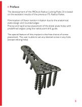
Preface The development of the PROlock Radius Locking Plate 2.0 is based on the excellent results of the previous ITS. Radius Plates. Minimization of flexor tendon irritation due to the anatomical plate design and rounded edges. Precise and rapid screw placement of the distal plate holes with predefined angles using the radiolucent drill guide. The special feature of this implant is the free choice of screw placement. The user is able to set any desired screw in any hole (except oblong hole).
Open the catalog to page 5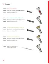
Spiral Drill, D=2.0mm, L=100mm, AO Connector Cortical Stabilization Screw, D=3.0mm, RH Spiral Drill, D=2.4mm, L=100mm, AO Connector Cancellous Stabilization Screw, D=3.0mm, RH Spiral Drill, D=2.0mm, L=100mm, AO Connector Spiral Drill, D=1.8mm, L=100mm, AO Connector Guide Wire, Steel, D=1.6mm, L=150mm, TR, w. thread
Open the catalog to page 6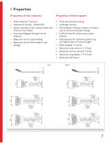
Properties Properties of the material: • Plate material: Titanium • Material of screws: TiAl6V4 ELI • Easier removal of the implant after the fracture has healed • Improved fatigue strength of the implant • Reduced risk of cold welding • Reduced risk of inflammation and allergy • Multi-directional locking • Left/right version • Minimization of flexor tendon irritation due to anatomical plate design • K-Wire holes for preliminary plate fixation • Oblong hole for optimal positioning and adjustment of radius length • Plate lenghts: 3, 5-hole • Optional wide version: 3, 5-hole • Optional narrow version:...
Open the catalog to page 7
♦ Predefined angles of the distal holes Version left Version right Hole number Direction proximal/distal Direction ulnar/radial
Open the catalog to page 8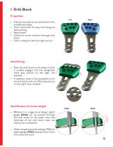
Drill Block Properties: • Precise and rapid screw placement with predefined angles • Click mechanism for easy mounting and dismounting • Radiolucent • Allows for screw insertion through drill block • Color coding for left and right version Assembling: • Slide the drill block to the distal end till it audible engages into the designated holes (see picture on the right, red marked) • In addition, there is the possibility to fix the drill block with a K-Wire (see picture on the right, blue marked) Identification of screw length: • PROlock II in single-hand design depth gauge (59026) can be pushed...
Open the catalog to page 9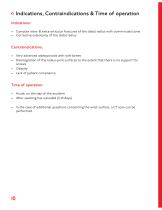
Indications, Contraindications & Time of operation Indications: • Complex intra- & extra-articular fractures of the distal radius with comminuted zone • Corrective osteotomy of the distal radius Contraindications: • Very advanced osteoporosis with soft bones • Disintegration of the radius-joint surfaces to the extent that there is no support for screws • Obesity • Lack of patient compliance Time of operation: • Acute, on the day of the accident • After swelling has subsided (5-10 days) • In the case of additional questions concerning the wrist surface, a CT scan can be performed.
Open the catalog to page 10
Surgical Technique
Open the catalog to page 11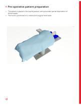
Pre-operative patient preparation • The patient is placed in the supine position with pneumatic partial deprivation of blood supply • The hand is positioned on a radiolucent surgical hand table
Open the catalog to page 12
Access The skin incision is performed on the distal forearm volar, above the tendon of the flexor carpi radialis and reaching to the crease of the wrist (FCR-approach). Split the deep fascia of the forearm. Release the pronator quadratus muscle from the distal radius beginning at the radial edge. Implantation Suspending the thumb with a counterpoise, the fracture is loosened and the length restored. The individual fragments are reduced with the appropriate instrumentation, and, if necessary, the comminuted zones are filled with bone substitute to achieve a provisional reduction in position and...
Open the catalog to page 13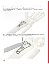
Possible temporary fixation of individual fragments using a guide wire, steel, D=1.6mm, L=150mm, TR w. Thrd. (35164-150) - check reduction under fluoroscopy. After anatomical reduction is achieved, the implant is chosen and, if required, its contour can be modified. First, fill the oblong hole with a D=2.7mm cortical screw (32271-XX) (suitable drills see page 6). Check the reduction and position of the plate under fluoroscopy.
Open the catalog to page 14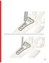
Due to the oblong hole it is possible to adjust the plate position either distal or proximal.
Open the catalog to page 15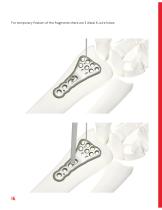
For temporary fixation of the fragments there are 3 distal K-wire holes.
Open the catalog to page 16
Next fill the shaft holes with either D=3.0mm cortical stabilization screws (37304-XX) or with D=2.7mm cortical screws (32271-XX) (suitable drills see page 6).
Open the catalog to page 17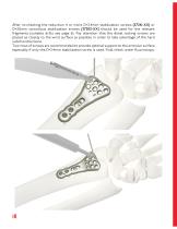
After re-checking the reduction 4 or more D=2.4mm stabilization screws (37241-XX) or D=3.0mm cancellous stabilization screws (37303-XX) should be used for the relevant fragments (suitable drills see page 6). Pay attention that the distal locking screws are placed as closely to the wrist surface as possible in order to take advantage of the hard subchondral bone. Two rows of screws are recommended to provide optimal support to the articular surface especially if only the D=2.4mm stabilization screw is used. Final check under fluoroscopy.
Open the catalog to page 18All I.T.S. catalogs and technical brochures
 CTN - Cannulated Tibia Nail
28 Pages
 ufs
ufs1 Page
 DHL
DHL2 Pages
 ITS
ITS2 Pages
 SCL
SCL32 Pages
 SLS
SLS24 Pages
 OL - Olecranon Locking Plate
24 Pages
 PHL
PHL24 Pages
 PHLs
PHLs20 Pages
 CLS
CLS28 Pages
 ACLS
ACLS20 Pages
 CFN
CFN32 Pages
 OLS
OLS24 Pages
 SR Sacral Rods
20 Pages
 HCS
HCS24 Pages
 TOS Twist-Off Screw
20 Pages
 TLS
TLS20 Pages
 PRS-RX
32 Pages
 HLS
HLS20 Pages
 ES
ES20 Pages
 SR
SR20 Pages
 FL
FL24 Pages
 PL - Pilon Locking Plate small
12 Pages
 FLS
FLS24 Pages
 PFL
PFL20 Pages
 DTL
DTL24 Pages
 HTO
HTO24 Pages
 PTL
PTL32 Pages
 DFL
DFL32 Pages
 CAS
CAS40 Pages
 FCN
FCN20 Pages
 HOL
HOL24 Pages
 CAL
CAL20 Pages
 DUL
DUL24 Pages


Archived catalogs


- Bone plate
- Compression plate
- Metallic compression plate
- Locking compression plate
- Distal compression plate
- Compression bone screw
- Metallic compression bone screw
- Proximal compression plate
- Arthrodesis nail
- Forearm compression plate
- Lateral compression plate
- Medial compression plate
- General purpose compression bone screw
- Tibia compression plate
- Metallic intramedullary nail
- Humerus compression plate
- Cannulated compression bone screw
- Radius compression plate
- Proximal fixation intramedullary nail
- Arthrodesis plate