


JEOL Application Data Sheet

JEOL Application Data Sheet
Cryo scanning electron microscopy (cryo SEM) is a technique used to image samples containing moisture without causing drying artifacts. It involves a cryo chamber for cleaving and coating frozen samples and a cold stage for imaging. The system uses a single liquid nitrogen tank to cool both the chamber and the stage, and includes a resistance heating vacuum evaporator for coating samples with gold and a heater for etching ice.
Operating Procedure
The procedure involves rapid freezing of the sample with liquid nitrogen before loading it into the cryo SEM. The sample is cleaved with a cold knife for internal structure imaging, etched to remove ice, and coated with gold for SEM imaging. Key steps include etching (ice sublimation) and preliminary freezing (physical fixation).
Etching Process
Etching controls the temperature of the frozen sample in vacuum, allowing ice to sublimate and providing information on water distribution. The sublimation temperature varies with the vacuum level in the SEM chamber and can be identified from the ice vapor pressure curve. An example is shown with oil-in-water emulsion, where etching reveals the distribution of ice.
Preliminary Freezing
Preliminary freezing, or physical fixation, involves freezing the sample with liquid nitrogen before loading it into the cryo chamber. Rapid freezing techniques, such as the metal contact method, are necessary to prevent sample structure damage due to ice crystal growth. This method involves pressing the sample onto a metal plate cooled with liquid nitrogen, resulting in clearer imaging of individual particles.
Conclusion
For optimal results, samples should be small and frozen rapidly to minimize ice crystal damage and ensure clear imaging.
Catalog excerpts
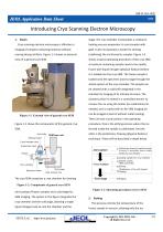
JEOL Application Data Sheet JE Introducing Cryo Scanning Electron Microscopy stage. The cryo chamber incorporates a resistance 1. Basics Cryo scanning electron microscopy is effective in heating vacuum evaporator to coat samples with imaging of samples containing moisture without gold. It also incorporates a heater for etching causing drying artifacts. Figure 1-1 shows an external (subliming) the ice formed in samples. Figure 1-3 view of a general cryo SEM. shows a typical operating procedure of this cryo SEM. A moisture containing sample needs to be rapidly frozen with liquid nitrogen (physical fixation) before it is loaded into the cryo SEM. The frozen sample is loaded onto the specimen process stage through the airlock system of the cryo chamber. The sample can be cleaved with a cold knife integrated in the chamber for imaging of its internal structure. The cleaved surface is etched in a controlled manner to remove the ice using the heater (ice sublimation) as needed, and is coated with Au for SEM imaging (or Figure 1-1. External view of general cryo SEM can be imaged at low kV without metal coating). There are two crucial points in the operating Figure 1-2 shows the components of this general cryo procedure. One is the etching process where the ice formed inside the sample is sublimated. And the Cold knife Etching heater other is the preliminary freezing (physical fixation) technique. These will be described in detail below. Liquid nitrogen tank Evaporator head Atmosphere Airlock Preliminary freezing (rapid freezing) (Freezing in atmosphere before loading sample into cryo chamber) Cryo chamber Cryo chamber Specimen holder Loading sample into cryo chamber Sample cutting (With cold knife) Etching (Sublimation of ice with heater) Coating Cold stage Cold stage Heat transfer line The cryo SEM comprises a cryo chamber for cleaving Figure 1-2. Components of general cryo SEM and coating of frozen samples and a cold stage for SEM imaging. The system in the figure integrates the cryo chamber and the cold stage, allowing a single liquid nitrogen tank to cool the chamber and the Figure 1-3. Operating procedure of cryo SEM 2. Etching This process controls the temperature of the frozen sample in vacuum, allowing only the ice Copyright(C) 2011 JEOL Ltd., All Rights Reserved
Open the catalog to page 1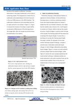
JEOL Application Data Sheet JE formed in the sample to sublimate. It provides information on the water distribution in the moisture 3. Notes on preliminary freezing containing sample. The temperature at which the ice Preliminary freezing is called physical fixation as sublimates varies depending on the level of vacuum opposed to chemical fixation. At the preliminary in the cryo SEM (pressure in the SEM chamber). The freezing process, a moisture containing sample is sublimation temperature can be identified from the frozen with liquid nitrogen before the sample is ice vapor pressure curve shown...
Open the catalog to page 2
JEOL Application Data Sheet JE Figure 3-2. Freezing by metal contact The sample was pressed onto a metal plate cooled down to the liquid nitrogen temperature for rapid freezing. Individual particles of polymer emulsion are visible. Figures 3-1 and 3-2 confirm that the ideal sample should be as small as possible and be frozen as fast as possible. Copyright(C) 2011 JEOL Ltd., All Rights Reserved
Open the catalog to page 3All Jeol catalogs and technical brochures
 Products Guide 2025
20 Pages
 Company Profile
20 Pages
Archived catalogs
 JCM-6000Plus
2 Pages
- Analysis software
- JEOL microscope
- JEOL laboratory microscope
- JEOL benchtop microscope
- Sample preparation system
- Visualization software
- Automatic sample processor
- Clinical chemistry analyzer
- Reporting software
- Windows software
- JEOL laboratory software
- Laboratory sample preparation system
- LED microscope
- Automated biochemistry analyzer
- Benchtop biochemistry analyzer
- JEOL spectrometer
- Benchtop sample preparation system
- Acquisition software
- Treatment software
- Measurement software