iFS

iFS
Catalog excerpts

iFS Advanced Femtosecond LaserA COMPREHENSIVE PLATFORM OF SURGICAL PRECISION INDICATIONS The iFS® Femtosecond Laser is an ophthalmic surgical laser indicated for use in patients undergoing surgery or treatment requiring initial lamellar resection of the cornea, in treatment requiring initial lamellar resection of the cornea to create tunnels for placement of corneal ring segments, in treatment requiring arcuate cuts/incisions in the cornea, penetrating and/or intrastromal, In lamellar IEK and corneal harvesting; in the creation of a corneal flap in patients undergoing LASIK surgery or other treatment requiring initial lamellar resection of the corneal, in the creation of a lamellar cut/resection of the cornea for lamellar IEK and for the creation of a penetrating cut/incision for penetrating IEK, in treatment requiring the creation of corneal channels for placement/ insertion of a corneal inlay device. LASIK FLAPS CUSTOMIZED CORNEAL INCISIONS LASER CATARACT INCISIONS CORNEAL CHANNEL CREATION INTRACORNEAL CUSTOMER SAFETY RING SEGMENTS EXPERIENCE TEAM INFORMATION
Open the catalog to page 1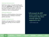
Built from a legacy of market leading and innovative IntraLase® Technology utilized in over 8 million procedures with more than 300 scientific citations, giving you confidence when treating patients PROVEN RESULTS Rooted in a solid foundation of precision and performance, helping to maximize patient outcomes exactly what it's supposed to do.” -JOHN VUKICH, MD PROVEN VERSATILITY Modular and upgradeable for multiple applications, giving you more surgical options CORNEAL CHANNEL INTRACORNEAL CUSTOMER SAFETY CREATION RING SEGMENTS EXPERIENCE TEAM INFORMATION
Open the catalog to page 2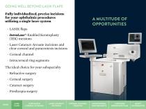
GOING WELL BEYOND LASIK FLAPS Fully individualized, precise incisions for your ophthalmic procedures utilizing a single laser system LASIK flaps • IntraLase®-Enabled Keratoplasty (IEK) incisions • Laser Cataract: Arcuate incisions and clear corneal and paracentesis incisions • Corneal channel • Intracorneal ring segments The ideal choice for your subspecialty • Refractive surgery • Corneal surgery • Cataract surgery • Presbyopia surgery CORNEAL CHANNEL INTRACORNEAL CUSTOMER SAFETY CREATION RING SEGMENTS EXPERIENCE TEAM INFORMATION
Open the catalog to page 3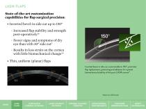
LASIK FLAPS State-of-the-art customization capabilities for flap surgical precision: I • nverted bevel-in side cut up to 150° • Increased flap stability and strength post-operatively1* • Fewer signs and symptoms of dry eye than with 30° side cut2 • Results in less strain on the cornea with little biomechanical change3,4 T • hin, uniform (planar) flaps Inverted bevel-in side cut, customizable to 150°, promotes flap replacement, positioning and adhesion for optimal biomechanical stability of the post-LASIK cornea1*. *Based on rabbit study LASIK FLAPS CUSTOMIZED CORNEAL INCISIONS LASER CATARACT...
Open the catalog to page 4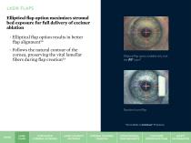
LASIK FLAPS Elliptical flap option maximizes stromal bed exposure for full delivery of excimer ablation • Elliptical flap option results in better flap alignment5,6 • Follows the natural contour of the cornea, preserving the vital lamellar fibers during flap creation5,6 Elliptical flap option available only with the iFS® Laser*. Standard round flap. *Not available on IntraLase® FS Systems LASIK FLAPS CUSTOMIZED CORNEAL INCISIONS LASER CATARACT INCISIONS CORNEAL CHANNEL CREATION INTRACORNEAL RING SEGMENTS CUSTOMER EXPERIENCE TEAM SAFETY INFORMAT
Open the catalog to page 5
Low energy setting, tight spot, and line separation: • Results in smooth stromal beds for easier flap lift7 • Helps minimize inflammatory tissue reaction8 • Fast flap creation7 Images of the iFS® Laser are provided by Melvin A. Sarayba, MD LASIK CUSTOMIZED LASER CATARACT CORNEAL CHANNEL INTRACORNEAL CUSTOMER SAFETY FLAPS CORNEAL INCISIONS INCISIONS CREATION RING SEGMENTS EXPERIENCE TEAM INFORMATION
Open the catalog to page 6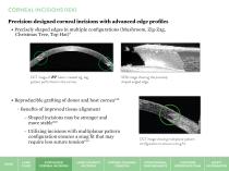
CORNEAL INCISIONS (IEK) Precision-designed corneal incisions with advanced edge profiles • Precisely shaped edges in multiple configurations (Mushroom, Zig-Zag, Christmas Tree, Top Hat)9 OCT image f iFS® Laser-created zig-zag o pattern performed on the cornea. SEM image howing the precisely s shaped angled edge. • Reproducible grafting of donor and host cornea9,10 • Benefits of improved tissue alignment – Shaped incisions may be stronger and more stable9,10 – Utilizing incisions with multiplanar pattern configuration ensures a snug fit that may require less suture tension9,10 LASIK FLAPS CUSTOMIZED...
Open the catalog to page 7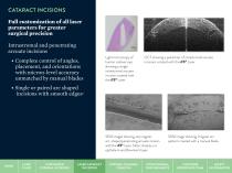
Full customization of all laser parameters for greater surgical precision Intrastromal and penetrating arcuate incisions • Complete control of angles, placement, and orientations with micron-level accuracy unmatched by manual blades • Single or paired arc shaped incisions with smooth edges11 Light microscopy of OCT showing a paired set of intrastromal arcuate human cadaver eye incisions created with the iFS® Laser. showing a single intrastromal arcuate incision created with the iFS® Laser. SEM images showing very regular, SEM image showing irregular arc arc-shaped penetrating arcuate...
Open the catalog to page 8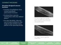
CATARACT INCISIONS Precision designed cataract incisions • Creation of multiplanar clear corneal and planar paracentesis incisions • Incisions have a precise construction with clear defined planes12 • Provides flexibility, allowing you to create incisions in your surgical suite and then move patient to the operating facility OCT side view image of the cornea after c reation of a triplanar clear corneal incision. N ote: incisions have a precise construction, w ith clear, defined planes. OCT image of 1-day post-surgical lear c corneal incision. LASIK FLAPS CUSTOMIZED CORNEAL INCISIONS LASER CATARACT...
Open the catalog to page 9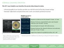
The iFS® Laser intuitive user interface for precise inlay channel creation • Advanced graphical user interface provides you with full control of the inlay channel settings • Parameter adjustments and patient data entry can be conveniently performed onscreen Multiple parameter setting options Superior to inferior size the channel: 3.6 mm-4.7 mm Distance between surgical field center and channel reference axis: 0.0 mm-2.8 mm Stromal depth of channel: 100-400 microns Distance between the channel reference axis and entry side cut: 3.7 mm-7.6 mm Angle from channel to epithelial surface: 30°-90° Note:...
Open the catalog to page 10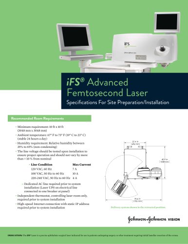

Archived catalogs
 whitestar signature pro
8 Pages
 IFS
IFS4 Pages
 COMPACT INTUITIV System
151 Pages
 iDesign - Refractive Studio
5 Pages
 The CATALYS® System
10 Pages
 LipiView® II
2 Pages
 COMPACT INTUITIV
5 Pages
 COMPACT INTUITIV
5 Pages
 ELLIPS® FX Technology.
6 Pages
 SOVEREIGN®
6 Pages
 WHITESTAR SIGNATURE® PRO
5 Pages


- Medimeas syringe
- Diode laser
- Drill
- IPL system
- Straight handpiece
- Safety syringe
- Intraocular lens
- Ophthalmic laser
- Surgery unit footswitch
- Surgery handpiece
- Table-top IPL system
- 0.5 ml syringe
- Floor-standing laser
- Monofocal intraocular lens
- Multifocal intraocular lens
- Dry eye diagnosis system
- Capsulotomy laser
- Meibography dry eye diagnosis system
- Ultrasonic handpiece
- Wireless footswitch