Dental Microscopes - general info

Dental Microscopes - general info
The document discusses the advantages and applications of dental microscopes in enhancing dental practices. It emphasizes the importance of precise vision in dental procedures and how microscopes can improve the quality of care.
Specifications and Features
The dental microscope offers a magnification range from x4 to x26, suitable for various dental applications. It is highlighted as an essential tool for procedures like micro-endodontic retreatment and surgical endodontic management.
Clinical Applications
Several clinical scenarios are presented, showcasing the use of microscopes in procedures such as apicectomy and root end filling. The document includes images demonstrating the detailed views achievable with magnification, such as the fitting of a crown and the examination of a resected root surface.
Expert Opinions
Quotes from dental professionals, including Dr. Charbel Allam and Prof. Gabriele Pecora, underline the significance of microscopes in modern dentistry. They stress that light and magnification have elevated the standard of dental care.
Conclusion
The document concludes by encouraging dentists to adopt microscopes in their practice to enhance precision and patient outcomes. It notes that technological advancements may lead to changes in features and specifications.
Catalog excerpts
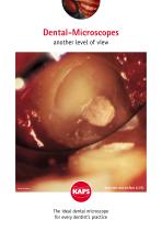
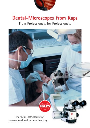
Dental-Microscopes another level of view Resected root surface (x 25) The ideal dental microscope for every dentist’s practice
Open the catalog to page 1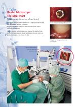
Dental Microscope: The ideal start The more you use, the more you will want to use it The need to maintain patients dentition for a longer period of time may lead to the use of advanced procedures: the micro-endodontic retreatment, the conventional and surgical endodontic management. Hex top of implant covered by soft tissue (16x) Training, practice, and technology have improved the quality of care provided to the population. Change your life and save your teeth, use dental microscopes ... but choose the best. Fitting of crown of upper central incisor on tooth (26x) View of a IRM retrofilling...
Open the catalog to page 2
The ML canal on a resected root of mandibular first molar (x 25) Isthmus between the two canals (x 25) Retromirror view of leakage on MB canal on a resected root of mandibular first molar (x 25) Retromirror view of retropreparation of MB and ML canal on a mandibular first molar (x 16) “Exact therapy requires exact vision.” Perfectly prepared canal with a ultrasonic tip (x25)
Open the catalog to page 3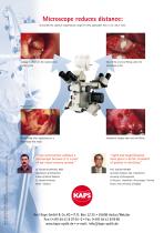
Microscope reduces distance: It provides the optimum magnification range for every application from x 4 to x 26 or more. Leakage is shown on the resected root View of the root end filling under the Clinical view of an apicectomy on a Endodontic Surgery after root end filling Layout: Grips Design, Rinker & Weber, Wetzlar Due to continual advances in technology, features and specifications are subject to change without notice. mandibular first molar “I can not practice without a microscope because it is a part of my visual sensory system.” “Light and magnification have given a better standard of...
Open the catalog to page 4All Karl Kaps catalogs and technical brochures
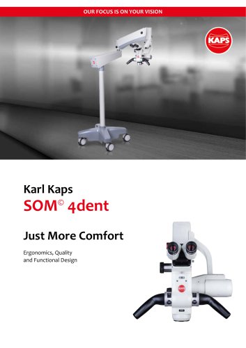 SOM4dent
12 Pages
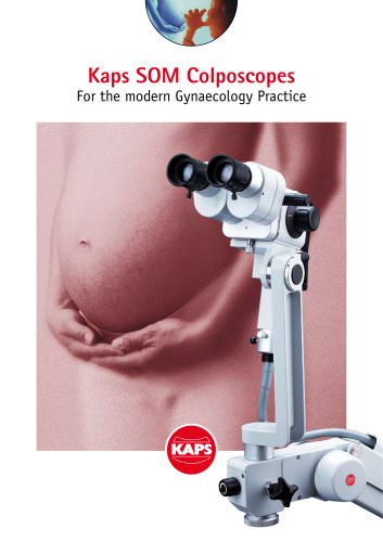
 Karl Kaps Colposcopes
16 Pages
 Kaps SOM® 62 Ophthal
8 Pages
 Kaps HD Video Camera
1 Page
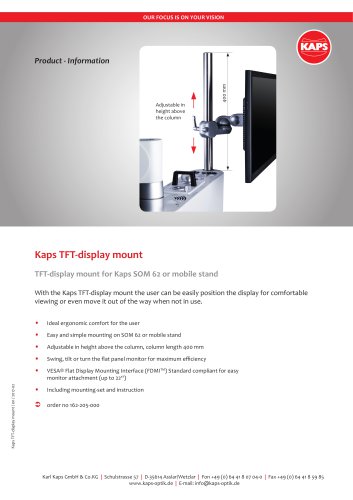 Kaps TFT display mount
2 Pages
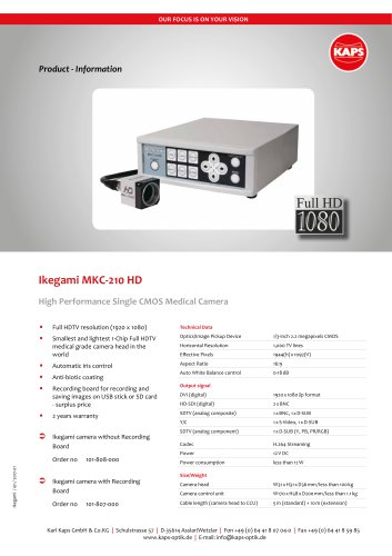 Kaps Ikegami
1 Page
 kaps operating microscope
2 Pages
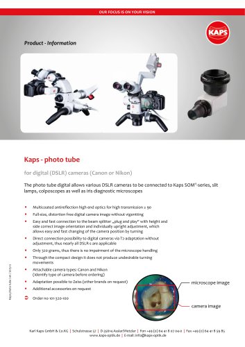 Kaps Photo Tube
1 Page
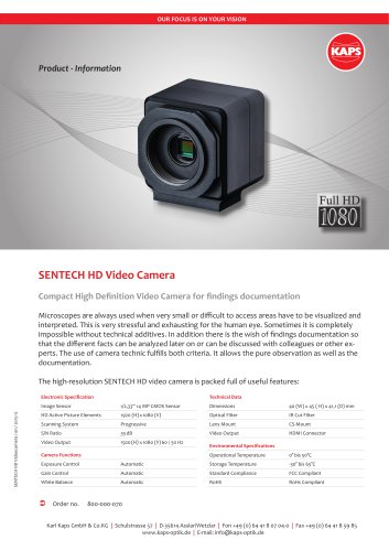
 SENTECH HD Video Camera
1 Page
 Kaps - Media Work Station BE
2 Pages


Archived catalogs
 SOM 62 Opthal
4 Pages
 Kaps Dental-Microscopes
6 Pages
 Videocolposcope ViCo
2 Pages
 Kaps KP 3000 Colposcopes
4 Pages
 SDM 60 Diagnosis Microscope
4 Pages
 Kaps SOM 42/52 Colposcopes
4 Pages
 Surgicalmicroscope SOM 62
4 Pages
 The KC Series
6 Pages
 SOM 62 for General Surgery
4 Pages
 Kaps SOM Handle bars
1 Page

- Stereo microscope
- Optical stereo microscope
- Slit lamp
- Table slit lamp
- Colposcope
- Benchtop stereo microscope
- Operating microscope
- Binocular stereo microscope
- Video colposcope
- Trolley-mounted colposcope
- LED stereo microscope
- Zoom stereo microscope
- Binocular colposcope
- Dental operating microscope
- Examination microscope
- Ophthalmic operating microscope
- Examination microscope on casters
- ENT operating microscope
- Medical stereo microscope
- ENT examination microscope