

MD3T™ Technique Overview
MD3T™ Technique Overview
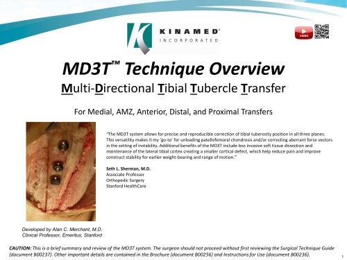
The MD3T™ system is designed for precise correction of tibial tuberosity position in multiple planes, offering benefits such as reduced soft tissue dissection and improved stability for early weight-bearing. It is particularly useful for addressing patellofemoral chondrosis and instability.
Traditional Tubercle Transfers
Traditional methods like the Elmslie-Trillat and AMZ have limitations, including risks of fractures and less control over transfer distances. The MD3T system addresses these issues with its innovative approach.
MD3T Basic Concept
The system uses two compound wedges (Primary & Secondary) to medialize the tibial tubercle. This allows for independent control of medial and anterior transfer distances.
System Benefits
The MD3T system offers flexibility, precision, safety, simplicity, and construct security. It allows for independent planning of medial and anterior transfers and supports accelerated rehabilitation.
Technique Overview
The procedure involves creating and transposing Primary and Secondary Wedges to achieve the desired tibial tubercle position. The technique allows for various transfer types, including medial, anteromedial, distal, and proximal.
Instruments and Procedure
The MD3T set includes guides and tools for precise bone cuts. Surgeons use their preferred fixation screws. The procedure involves attaching guides, making precise cuts, planning transfers, and securing the tubercle with screws.
Bone Void Filler Application
Fast-setting bone void filler is used to fill spaces created by bone resection, ensuring stability and proper positioning of the tubercle.
Conclusion
The MD3T system provides a comprehensive solution for tibial tubercle transfer, enhancing surgical outcomes through its innovative design and technique.
Catalog excerpts
MD3T™ Technique Overview Multi-Directional Tibial Tubercle Transfer For Medial, AMZ, Anterior, Distal, and Proximal Transfers “The MD3T system allows for precise and reproducible correction of tibial tuberosity position in all three planes. This versatility makes it my ‘go-to’ for unloading patellofemoral chondrosis and/or correcting aberrant force vectors in the setting of instability. Additional benefits of the MD3T include less invasive soft tissue dissection and maintenance of the lateral tibial cortex creating a smaller cortical defect, which help reduce pain and improve construct stability for earlier weight-bearing and range of motion.” Seth L. Sherman, M.D. Associate Professor Orthopedic Surgery Stanford HealthCare Developed by Alan C. Merchant, M.D. Clinical Professor, Emeritus, Stanford CAUTION: This is a brief summary and review of the MD3T system. The surgeon should not proceed without first reviewing the Surgical Technique Guide (document B00237). Other important details are contained in the Brochure (document B00256) and Instructions for Use (document B002
Open the catalog to page 1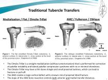
Traditional Tubercle Transfers Medialization / Flat / Elmslie-Trillat The Elmslie-Trillat is a straight medialization (without anteriorization) that is performed for correction of patellar instability and lateral patellar compression syndrome with no or minimal chondrosis. The Elmslie-Trillat may have a thin bone shingle and small proximal buttress, which can lead to fracture or pull-off. The AMZ creates a large cortical defect with a known risk of proximal tibial fracture. The slope of the AMZ bone resection controls both anterior and medial transfer distance
Open the catalog to page 2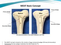
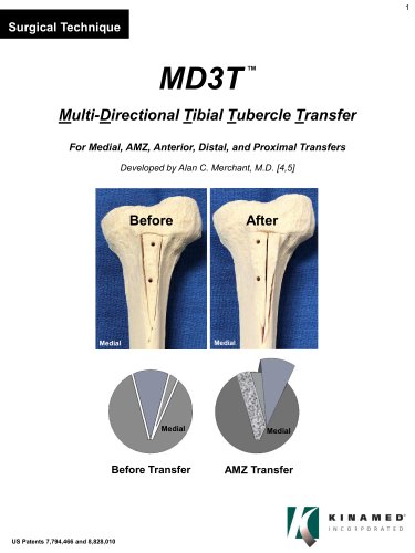
MD3T Basic Concept Primary Wedge Primary Wedge Secondary Wedge Secondary Wedge All anatomical photos in this Guide represent a Left Tibia The MD3T concept is based on the creation of two compound wedges (Primary & Secondary). Transposing the two wedges medializes the tibial tubercle (TT).
Open the catalog to page 3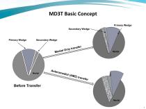
MD3T Basic Concept Primary Wedge Secondary Wedge Primary Wedge Secondary Wedge Before Transfer
Open the catalog to page 4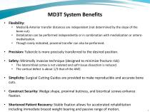
MD3T System Benefits • Flexibility: • Medial & Anterior transfer distances are independent (not determined by the slope of the bone cut). • Distalization can be performed independently or in combination with medialization or antero-medialization. • Though rarely indicated, proximal transfer can also be performed. • Precision: Tubercle is more precisely transferred to the desired position. • Safety: Minimally invasive technique (designed to minimize fracture risk) • The lateral tibial cortex is not violated and soft tissue dissection is reduced. • The cortical defect is about 1/3 that of the AMZ....
Open the catalog to page 5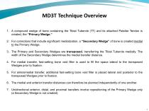
MD3T Technique Overview 1. A compound wedge of bone containing the Tibial Tubercle (TT) and its attached Patellar Tendon is created, the “Primary Wedge.” 2. For corrections that include significant medialization, a “Secondary Wedge” of bone is created medial to the Primary Wedge. 3. The Primary and Secondary Wedges are transposed, transferring the Tibial Tubercle medially. The width of the Secondary Wedge determines the medial transfer distance. 4. For medial transfer, fast-setting bone void filler is used to fill the space lateral to the transposed Wedges prior to fixation. 5. For anteromedial...
Open the catalog to page 6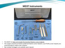
** « Multidirectional Tibial Tubercle Transfer System • The MD3T set does not include the bicortical fixation screw implants. • The surgeon should use his/her preferred fixation screw implants. Low Profile screw implants are recommended to reduce skin irritation. • Non-slotted Outriggers are availab
Open the catalog to page 7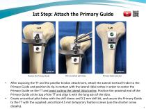
1st Step: Attach the Primary Guide Left Lateral Position the Primary Guide Lateral Drill unicortical pilot holes Primary Guide secured After exposing the TT and the patellar tendon attachment, attach the Lateral Cortical Finder to the Primary Guide and position its tip in contact with the lateral tibial cortex in order to center the Primary Guide on the TT and avoid cutting the lateral tibial cortex. Position the proximal end of the Primary Guide at the top of the TT and align it with the long axis of the tibia. Create unicortical pilot holes with the drill sleeve and 3.2 mm drill bit, and secure...
Open the catalog to page 8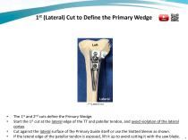
1st (Lateral) Cut to Define the Primary Wedge The 1st and 2nd cuts define the Primary Wedge. Start the 1st cut at the lateral edge of the TT and patellar tendon, and avoid violation of the lateral cortex. Cut against the lateral surface of the Primary Guide itself or use the Slotted Sleeve as shown. If the lateral edge of the patellar tendon is exposed, lift it up to avoid cutting it with the saw blad
Open the catalog to page 9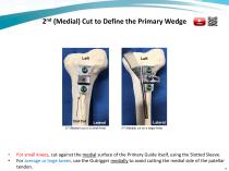
2nd (Medial) Cut to Define the Primary Wedge Replace with new Fig 6 from B00237 Lateral Lateral 2nd (Medial) cut on a small knee Lateral 2nd (Medial) cut on a larger knee For small knees, cut against the medial surface of the Primary Guide itself, using the Slotted Sleeve. For average or large knees, use the Outrigger medially to avoid cutting the medial side of the patellar tendon. 1
Open the catalog to page 10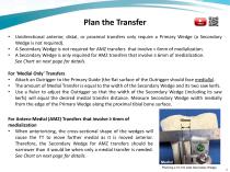
Unidirectional anterior, distal, or proximal transfers only require a Primary Wedge (a Secondary Wedge is not required). A Secondary Wedge is not required for AMZ transfers that involve < 6mm of medialization. A Secondary Wedge is only required for AMZ transfers that involve ≥ 6mm of medialization. See Chart on next page for details. For ‘Medial Only’ Transfers • Attach an Outrigger to the Primary Guide (the flat surface of the Outrigger should face medially). • The amount of Medial Transfer is equal to the width of the Secondary Wedge and its two saw kerfs. • Use a Ruler to adjust the Outrigger...
Open the catalog to page 11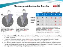
Planning an Anteromedial Transfer Planned Medial Transfer (mm) Planned Anterior Transfer (mm) Medial For 10mm medial & 5mm anterior, width of Secondary Wedge should be 7mm. See Table. For 10mm medial & 10mm anterior, width of Secondary Wedge should be 5mm. See Table. Width of Secondary Wedge (mm) To achieve the planned AMZ transfer, create a Secondary Wedge (including saw kerfs) having the proximal width shown in red italics. Note: For smaller medial transfers, a Secondary Wedge is not needed, as shown by the asterisks (*). For Anteromedial Transfers, the shape of the Primary Wedge causes the...
Open the catalog to page 12All Kinamed catalogs and technical brochures
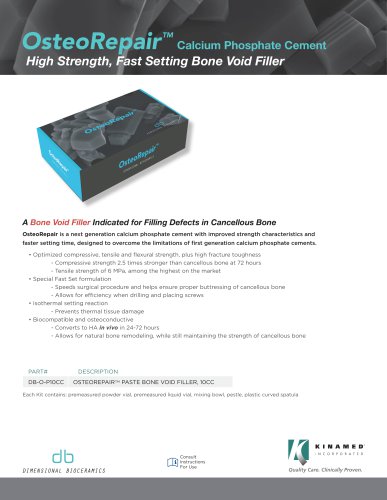 OsteoRepair
2 Pages
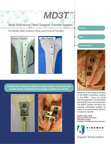 MD3T
2 Pages
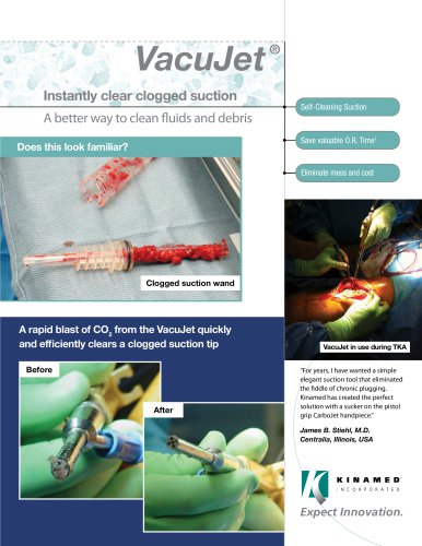 VacuJet ®
2 Pages
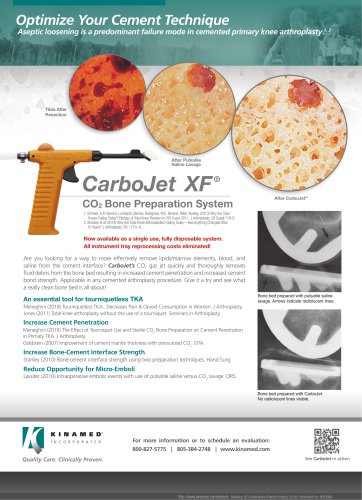
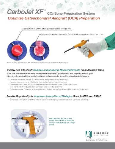
 CarboJet XF ®
2 Pages
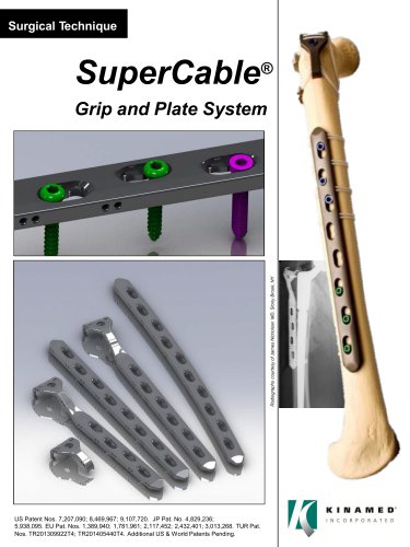
 SuperCable
4 Pages
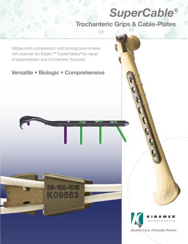
 SuperCable Brochure
4 Pages
 SuperCable Flyer
1 Page
 KineMatch® PFR
4 Pages


- Bone plate
- Compression plate
- Metallic compression plate
- Locking compression plate
- Titanium compression plate
- Proximal compression plate
- Surgical navigation system
- Optical surgical navigation system
- Cranial fixation system
- Non-absorbable cranial fixation system
- Pulsed lavage system
- Bone cerclage wiring
- Orthopedic surgery pulsed lavage system
- Orthopedic surgery navigation system
- Multifilament polymer bone cerclage
- Sternal closure bone cerclage wiring
- Patello-femoral prosthesis