

Multi-Directional Tibial Tubercle Transfer
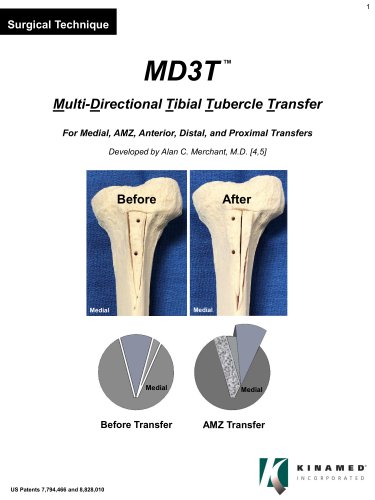
Multi-Directional Tibial Tubercle Transfer
The Multi-Directional Tibial Tubercle Transfer (MD3T) is a surgical technique aimed at treating patellofemoral disorders by transferring the tibial tubercle (TT). This method allows for precise movement of the TT in multiple directions, minimizing the risk of tibial fracture. It is particularly beneficial for patients with recurrent patellar dislocation, minimal patellar chondrosis, and tibial tubercle lateralization.
The MD3T System is suitable for patients who have reached skeletal maturity and require tibial tubercle transfer. It can address various causes of patellofemoral disorders and is applicable for medial transfer alone or combined with anterior transfer for anteromedialization (AMZ).
- Attach the Primary Guide: A vertical incision is made over the TT, and the Primary Guide is aligned with the tibia's long axis, secured with temporary fixation screws.
- Planning the Correction: Preoperative planning determines the transfer direction and magnitude, with the Secondary Wedge dictating the medial transfer distance.
- Make the 1st and 2nd Cuts: These cuts define the Primary Wedge, including the TT and its attached patellar tendon.
- Plan the Transfer & Make the 3rd Cut: A Secondary Wedge is required for AMZ transfers involving significant medialization.
- Make the Proximal Cut and Free the Wedge(s): The proximal cut frees the wedges for transposition.
- Transpose the Wedges: The wedges are repositioned to adjust the TT, with intraoperative verification of the correction.
- Finalize the Transfer, Prepare and Apply the Bone Void Filler: Bone void filler is applied around the wedges to secure their position.
- Secure the Fixation: Final pilot holes are drilled, and fixation is secured with caution to avoid neurovascular damage.
The MD3T technique provides a comprehensive approach to patellofemoral disorders, allowing for tailored surgical interventions based on individual patient needs, enhancing outcomes and reducing complications.
A pilot hole is drilled using a 3.2 mm drill bit, ensuring it passes through the posterior cortex. Instruments from the preferred screw implant set are used to over-drill the pilot hole, creating a lag effect and countersinking the anterior opening. A depth gauge selects the appropriate length for a fixation screw implant, which is inserted until it catches the posterior cortex. The process is repeated for the distal hole, and screws are tightened alternately. Additional security can be achieved with a third fixation screw implant. Sharp or protruding edges are rounded off to avoid impinging on the patellar tendon.
For various transfer types, bone void filler is applied to compensate for resected bone and achieve the desired correction. For unidirectional Anterior Transfer, filler is placed lateral to the Primary Wedge. For Medial Transfer, filler is placed lateral to the bone Wedge(s). For unidirectional Distal Transfer, the desired distance is planned using preoperative images. For Antero-Medial (AMZ) Transfer, filler is placed both laterally and posteriorly under the bone Wedge(s). For unidirectional Proximal Transfer, bone is resected at the proximal end of the Primary Wedge, and filler is placed laterally.
The MD3T system includes components such as the Primary Guide, Lateral Cortical Finder, Set Screw, Driver, Drill Sleeve, Drill, Temporary Fixation Screws, Primary Guide Sleeve, Outrigger Saw Guides, Osteotomes, Ruler, Steinmann Pin, Autoclave Tray, and MD3T OsteoRepair™ Paste Bone Void Filler. It also mentions required and recommended items not included in the set, such as Fast Setting Bone Void Filler.
Catalog excerpts
Surgical Technique Multi-Directional Tibial Tubercle Transfer For Medial, AMZ, Anterior, Distal, and Proximal Transfers Developed by Alan C. Merchant, M.D. [4,5] Before Transfer
Open the catalog to page 1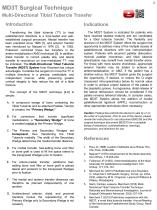
MD3T Surgical TechniqueMulti-Directional Tibial Tubercle TransferIntroduction Transferring the tibial tubercle (TT) to treat patellofemoral disorders is a time-tested and well-established surgical procedure. Medial transfer has been practiced since 1888 [1], and anterior transfer was introduced by Maquet in 1976 [2]. In 1983, Fulkerson combined these two transfers in the antero-medialization (AMZ) technique [3]. In addition, distal transfer to correct patella alta, and lateral transfer to reconstruct an over-medialized TT, may be indicated. The Multi-Directional Tibial Tubercle Transfer (MD3T)...
Open the catalog to page 2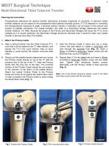
MD3T Surgical Technique Multi-Directional Tibial Tubercle Transfer Planning the Correction Preoperatively determine the desired transfer direction(s) including magnitude of correction. A planned medial transfer distance can be based on the preoperative tibial tubercle–trochlear groove (TT-TG) distance or correcting the intraoperatively measured Q angle. A planned anterior transfer, if indicated, can be based on the amount and location of patellar chondrosis. For medial transfers, the width of a Secondary Wedge determines the medial transfer distance. For AMZ, because the shape of the Primary...
Open the catalog to page 3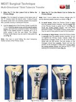
MD3T Surgical Technique Multi-Directional Tibial Tubercle Transfer 2. Make the 1st Cut (the Lateral Cut) to Define the Primary Wedge Caution: The 1st (lateral) cut begins at the lateral side of the TT, staying inside the lateral cortex. Cutting through the lateral cortex here can severely weaken the proximal tibia, possibly causing a subsequent fracture. Note: Cuts 1 and 2 define the Primary Wedge (the TT and its attached patellar tendon) as shown in Fig. 5. A. Use the lateral side of the Primary Guide to guide the 1st Cut. Avoid the lateral cortex and the lateral patellar tendon, and work from...
Open the catalog to page 4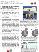
MD3T Surgical Technique Multi-Directional Tibial Tubercle Transfer 4. Plan the Transfer & Make the 3rd Cut to Define the Secondary Wedge, if necessary Note: Unidirectional anterior, distal, or proximal transfers only require a Primary Wedge (a Secondary Wedge is not necessary). A Secondary Wedge is only required for AMZ transfers that involve ≥ 6mm of medialization. A. For unidirectional anterior, distal, proximal, or combined transfers, a Secondary Wedge is not required. Skip to Step 5. B. For Antero-Medial (AMZ) transfers that involve < 6mm of medialization, a Secondary Wedge is not required....
Open the catalog to page 5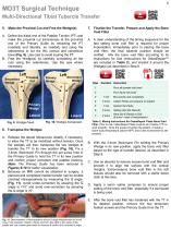
MD3T Surgical TechniqueMulti-Directional Tibial Tubercle Transfer5. Make the Proximal Cut and Free the Wedge(s)7. Finalize the Transfer, Prepare and Apply the Bone Void Filler A. Define the distal end of the Patellar Tendon (PT) and make the proximal cut transversely at the proximal end of the Primary Guide by retracting the PT (medially and laterally, as needed) and using the osteotomes to cut the thin cortical and cancellous bone (Fig. 9). Use care to avoid injuring the PT. B. Free the Wedge(s) by carefully completing all the cuts using the osteotomes. Use the saw where necessary. A. A clear...
Open the catalog to page 6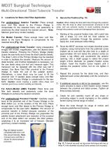
MD3T Surgical Technique Multi-Directional Tibial Tubercle Transfer 8. Locations for Bone Void Filler Application For unidirectional Anterior Transfer: Place enough bone void filler lateral to the Primary Wedge to compensate for the resected bone, and posteriorly to anteriorize the tibial tubercle the desired amount. Note: Maintain good bony contact medially. For Medial Transfer: Place enough bone void filler lateral to the bone Wedge(s) to compensate for the resected bone (Fig. 12). For unidirectional Distal Transfer: Using preoperative images with known magnification, plan the desired distal...
Open the catalog to page 7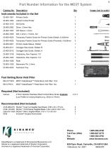
23-800-1002 Lateral Cortical Finder 1 23-800-4520 Temporary Fixation Screw for Primary Guide (Distal), 4.5x20mm 2 23-800-4526 Temporary Fixation Screw for Primary Guide (Proximal), 4.5x26mm 2 23-800-2001 Primary Guide Sleeve, Slotted 1 23-800-2011 Outrigger Saw Guide, Slotted, B 1 23-800-2013 Outrigger Saw Guide, Slotted, A 1 Fast Setting Bone Void Filler DB-O-P10CC MD3T OsteoRepair™ Paste Bone Void Filler, 10cc DB-O-P5CC MD3T OsteoRepair™ Paste Bone Void Filler, 5cc Various 4.5mm diameter Stainless Steel Cortical Bone Screw Implants 2 or 3 (Low Profile) of varying lengths (e.g. 25mm to 70mm)...
Open the catalog to page 8All Kinamed catalogs and technical brochures
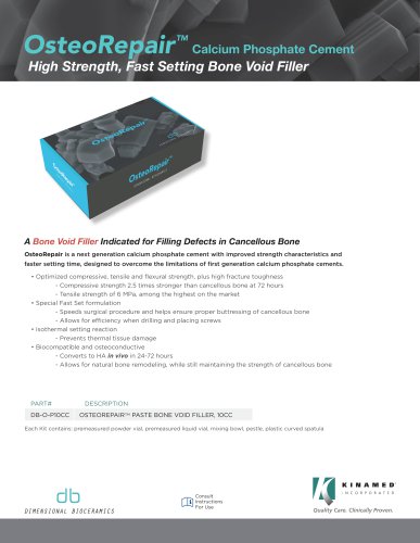 OsteoRepair
2 Pages
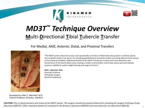 MD3T™ Technique Overview
21 Pages
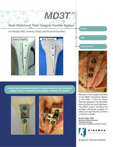 MD3T
2 Pages
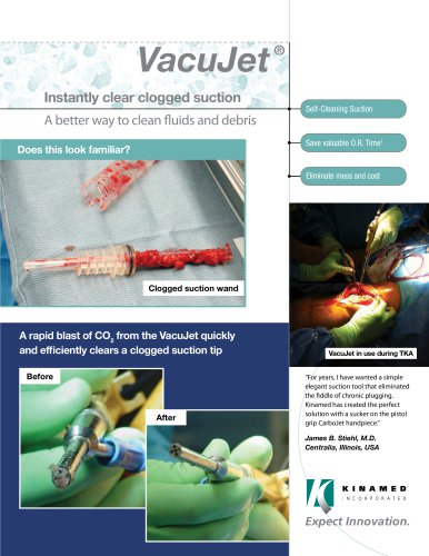 VacuJet ®
2 Pages
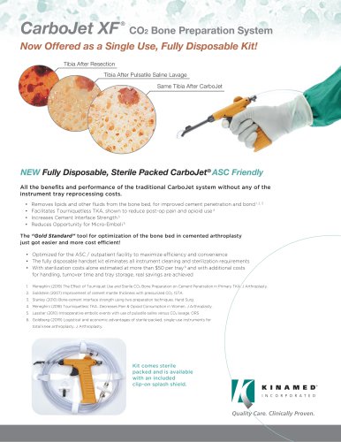 CarboJet XF ®
2 Pages
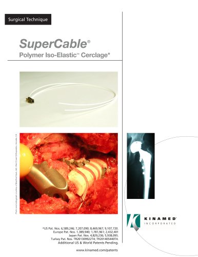 SuperCable
4 Pages
 SuperCable Brochure
4 Pages
 SuperCable Flyer
1 Page
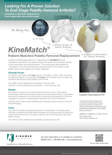
 KineMatch® PFR
4 Pages


- Bone plate
- Compression plate
- Metallic compression plate
- Locking compression plate
- Titanium compression plate
- Proximal compression plate
- Surgical navigation system
- Optical surgical navigation system
- Cranial fixation system
- Non-absorbable cranial fixation system
- Pulsed lavage system
- Bone cerclage wiring
- Orthopedic surgery pulsed lavage system
- Orthopedic surgery navigation system
- Multifilament polymer bone cerclage
- Sternal closure bone cerclage wiring
- Patello-femoral prosthesis