Catalog excerpts

Digital Variance Angiography a new era begins in radiation protection
Open the catalog to page 1
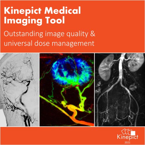
KINEPICT MAKES X-RAY ANGIOGRAPHY SAFER AND MORE POWERFUL Kinepict Health Ltd. has the vision to revolutionize x-ray angiography. We provide an advanced image analysis solution called Digital Variance Angiography implemented in the Kinepict Medical Imaging Tool: X-Safe that multiplies the abilities of your angiograhy suite. Summer 2020
Open the catalog to page 2
RADIATION PROTECTION • Despite the protective solutions used in modern healthcare the demand for radiation protection measures are higher than ever (Miller et al. Cardiovasc Intervent Radiol, 2010, 33.2, 230–239; Santos et al. Eur J Vasc Endovasc Surg, 2020, 59.4:654-660; Roguin et al. Am J Cardiol, 2013,111.9:1368-72) Today DSA represents 60-90% of the total radiation load in endovascular interventions The principle is to keep doses As Low As Reasonably Practicable (ALARP). This is achieved by various combinations of three basic parameters: time of exposure, distance from the source and...
Open the catalog to page 3
DIGITAL VARIANCE ANGIOGRAPHY • A platform independent x-ray image processing technology called Digital Variance Angiography (DVA) is available in the Kinepict Medical Imaging Tool: XSafe Tested and validated in clinical studies [1-4]: DVA provides better image quality at lower frame rates (1-2 FPS instead of 4-6 FPS). Instant image generation and adjustment, no post-processing needed. Peer-reviewed articles [1] Gyánó et al. Radiology, 2019, 290.1: 246-253. [2] Óriás et al. Investigative radiology, 2019, 54.7: 428-436. [3] Gyánó et al. Cardiovasc Intervent Radiol, 2020, 43:1226-1231. [4]...
Open the catalog to page 4
IMAGE QUALITY COMPARISON Radiation protection in ICM angiography Comparison of representative normal dose (1.2 µGy/frame) DSA and low-dose (0.36 µGy/frame) DVA images obtained in the abdominal (left) and femoral (right) regions. The corresponding image pairs were taken from the same patient and same direction in two consecutive runs, but the different regions belong to different patients. Brightness/contrast adjustments and pixel shift were applied to DSA and DVA images using the Siemens Syngo and the Kinepict workstation, respective
Open the catalog to page 5
DAILY ROUTINE USE OF DVA TECHNOLOGY Common femoral artery and femoro-popliteal multiplex angioplasty using DVA technology at the Heart and Vascular Center of Semmelweis University KINEPICT ACADEMY • CO2 angiography live case • Introduction to the software and DVA image types • DVA and DSA image quality comparison • Kinepict’s integration into the workflow
Open the catalog to page 6
AVAILABLE FUNCTIONS AFTER AUTOMATIC DVA GENERATION Kinepict Medical Imaging Tool: X-Safe • • • CE Certificate Nr.: HD 60134971 001 FDA approved medical device (510(K) Nr.: K190993) Validated and used daily in clinical environm
Open the catalog to page 7
www.kinepict.com Szabolcs Osváth, Ph.D. (CEO) Júlia utca 11. Budapest, 1026-H Reference sites and research collaborations Over 3 500+ patients and 50 000+ DVA images • Innsbruck Homburg • Salzburg Marburg Frankfurt Nuremberg Aachen • Rome
Open the catalog to page 8All Kinepict Health Kft catalogs and technical brochures
-
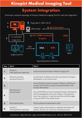 System integration
1 Pages
-
 Leaflet
2 Pages
-
 CO2 brochure
8 Pages