Catalog excerpts

Digital Variance Angiography a Paradigm Shift in CO2 Angiography
Open the catalog to page 1
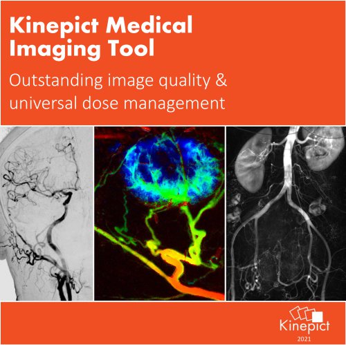
KINEPICT MAKES X-RAY ANGIOGRAPHY SAFER AND MORE POWERFUL Kinepict Health Ltd. is a start-up with the vision to revolutionize x-ray angiography. We provide an advanced image analysis solution called Digital Variance Angiography implemented in the Kinepict Medical Imaging Tool: X-Safe that multiplies the abilities of CO2 angiography. Summer 2020
Open the catalog to page 2
… was introduced as a negative contrast agent in medical imaging decades ago … can be used safely in patients with impaired renal function and with history of contrast induced nephropathy, a major cause of acute renal failure in hospitalized patients (Dam et al., Neth J Med, 2008, 66(10), 416-22.) … can replace ICM in wide variety of procedures like balloon angioplasty and stenting of arteries in the lower extremities, aortic aneurism repairs, aorography, renal and prostatic artery angiography, inferior vena cava and portal vein imaging, splenoportography, tumor embolization procedures,...
Open the catalog to page 3
DIGITAL VARIANCE ANGIOGRAPHY • Kinepict’s x-ray image processing technology called Digital Variance Angiography (DVA) is available in the Kinepict Medical Imaging Tool: X-Safe Tested and validated in clinical studies: DVA provides higher signal-to-noise ratio and better image quality than DSA in lower limb angiography even with ICM and CO2 [1-3] Visual comparison: the percent of DVA preference over DSA by regions (Al: abdominal-iliac; FP: femoral-popliteal; CT: crural-talar) from the BácsKiskun County Hospital (BKCH) and The Heart and Vascular Center of Semmelweis University (HVC) [2]...
Open the catalog to page 4
IMAGE QUALITY COMPARISON Case 1.: CO2 angiogram of a high-degree femoro-popliteal stenosis and occlusion. On post-dilatation image DVA clearly shows the non-significant residual stenosis also (1 FPS with Siemens Evenflow preset) [3]. Case 2.: High-grade stenosis on the left external iliac artery. After the first ballooning, an instable residual stenosis remained that is clearly visible on the DVA image (3 FPS with Siemens Evenflow preset) [3].
Open the catalog to page 5
DAILY ROUTINE USE OF DVA TECHNOLOGY CO2 angiography of the femoropopliteal region, balloon angioplasty, and self-deploying stent implantation at the Heart and Vascular Center of Semmelweis University KINEPICT ACADEMY • CO2 angiography live case • Introduction to the software and DVA image types • DVA and DSA image quality comparison • Kinepict’s integration into the workflow EPISODES AVAILABLE
Open the catalog to page 6
AVAILABLE FUNCTIONS AFTER DVA GENERATION Kinepict Medical Imaging Tool: X-Safe • • • CE Certificate Nr.: HD 60134971 001 FDA approved medical device (510(K) Nr.: K190993) Validated and used daily in clinical environm
Open the catalog to page 7
www.kinepict.com Szabolcs Osváth, Ph.D. (CEO) Júlia utca 11. Budapest, 1026-H Reference sites and research collaborations Over 3 500+ patients and 50 000+ DVA images • Innsbruck Homburg • Salzburg Marburg Frankfurt Nuremberg Aachen • Rome
Open the catalog to page 8All Kinepict Health Kft catalogs and technical brochures
-
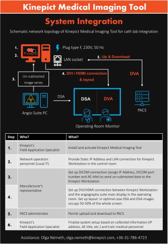 System integration
1 Pages
-
 Leaflet
2 Pages
-
 70% X-ray dose reduction
8 Pages