
Catalog excerpts

Kinepict Medical Imaging Tool Outstanding image quality & universal dose management
Open the catalog to page 1
KINEPICT MAKES X-RAY ANGIOGRAPHY SAFER AND MORE POWERFUL Kinepict Health Ltd. has the vision to revolutionize x-ray angiography. We provide an advanced image analysis solution called Digital Variance Angiography implemented in the Kinepict Medical Imaging Tool: X-Safe that multiplies the abilities of your angiograhy suite. Summer 2020
Open the catalog to page 2
KINEPICT MEDICAL IMAGING TOOL • … can visualize blood vessel structures by detecting the movement of the contrast medium bolus in standard-of-care angiography examinations. The platform independent software has provided safer and faster interventions for many thousand patients so far in our reference site network worldwide. Charachteristics that make the software unique for interventional radiologists Digital Variance Angiography Automatic image generation, display and adjustment Seamless integration, secure file handling, transfer to and from PACS Dual user interface: operating room and...
Open the catalog to page 3
DIGITAL VARIANCE ANGIOGRAPHY I. Digital Variance Angiography (DVA) images are generated from the unsubtracted X-ray image series by calculating the standard deviation of intensity values for every pixel. The proprietary algorithm of Kinepict enhances the contrast media-related visual information and suppresses the noise, thereby the image quality is greatly improved. This quality reserve assures universal dose management capabilities for DVA. Pixels of the original angio series MASK SUBTRACTION KINEPICT PROPRIETARY MINIMUM OR AVERAGE INTENSITY Left: DSA and DVA side-by-side comparison....
Open the catalog to page 4
DIGITAL VARIANCE ANGIOGRAPHY II. • DVA is implemented in the Kinepict Medical Imaging Tool software Instant image, video generation and adjustment, no post-processing needed. Tested and validated in clinical studies [1-5]: DVA provides higher contrast-tonoise ration and better image quality [1,4,5]. These benefits are achieved at lower frame rates (1-2 FPS instead of 46 FPS) in CO2 angiography [2,3]. Peer-reviewed articles [1] Gyánó et al. Radiology, 2019, 290.1: 246-253. [2] Óriás et al. Invest.Radiol., 2019, 54.7: 428-436. [3] Gyánó et al. CVIR, 2020, 43:1226-1231. [4] Bastian et al....
Open the catalog to page 5
DIGITAL VARIANCE ANGIOGRAPHY III. On color-coded DVA (ccDVA) images and videos the coloring represents the arrival latency of the contrast media according to the color spectrum of visible light. Examples of ccDVA images:
Open the catalog to page 6
Automatic generation, display and adjustment I. The DVA image is generated, adjusted and displayed in less than 2 seconds. Manual image manipulation is also available. Image adjustment functions: • • Color coding start & end (automatic) Pixelshift – motion artifact removal Invert colors (automatic) Distance compare Add coloring to selected area Original (left) and inverted (right) DVA images Brightness and Contrast (automatic) Undo & redo Almost all functions can be personalized at the Personal Preferences menu. You will receive image and video type
Open the catalog to page 7
Automatic generation, display and adjustment II. Adjustment options are also available from the operating room with an air-mouse. Left column: • • • • • • Active adjustment tool Cursor location pointer Image and video type selection Rollback image – display a previously selected image (e.g. pre-dilatation angio) Roadmap Set opacity of roadmap Right column: • • • • • Brightness & Contrast Pixelshift Fixed ROI Pixelshift Inve
Open the catalog to page 8
Seamless integration, secure file handling, transfer to and from PACS Files can be received directly from the angiography suite main computer, from PACS servers or from a desktop user directory. The generated files are sent directly to the PACS and saved to the desired directory. Un-subtracted image series Tested with • Philips Allura Clarity Siemens Artis Pheno Philips Allura Xper File handling options Operating room Download from PACS (automatic) Export DVA as DICOM, image or text Upload to PACS (automatic) Delete received files
Open the catalog to page 9
Dual user interface: operating room and workspace The Kinepict Medical Imaging Tool is designed in order to minimize the need of manual work to operate and to occupy almost zero workload of the medical staff. Control room (left) and operating room view (right), 2 screens are active simultaneously. Certificate Nr.: HD 60134971 001 FDA approved medical device (510(K) Nr.: K190993) Validated and used daily in clinical environm
Open the catalog to page 10
DAILY ROUTINE USE OF DVA TECHNOLOGY Common femoral artery and femoro-popliteal multiplex angioplasty using DVA technology at the Heart and Vascular Center of Semmelweis University KINEPICT ACADEMY • CO2 angiography live cases • Introduction to the software and DVA image types • DVA and DSA image quality comparison • Kinepict’s integration into the workflow
Open the catalog to page 11
www.kinepict.com BUSINESS INQUIRIES TECHNOLOGY & INSTALLATION Júlia utca 11. Budapest, 1026-H Reference sites and research collaborations Over 3 500+ patients and 50 000+ DVA images • Innsbruck Homburg • Salzburg Marburg Frankfurt Nuremberg Aachen • Rome
Open the catalog to page 12All Kinepict Health Kft catalogs and technical brochures
-
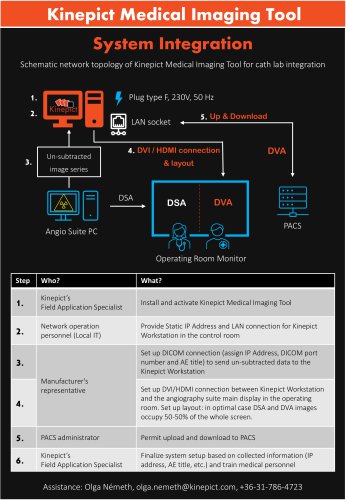 System integration
1 Pages
-
 Leaflet
2 Pages
-
 CO2 brochure
8 Pages
-
 70% X-ray dose reduction
8 Pages