- Catalogs
- Kowa Medical Care
- Nonmyd WX3D
Nonmyd WX3D

Nonmyd WX3D
- Simultaneous stereo 3D and 2D imaging with quick capture.
- Easy operation, reducing operator time and increasing patient throughput.
- Effective for assessing and monitoring glaucoma, with accurate verification of the cup and disc's physiological state.
- Advanced networking and connectivity for efficient workflow.
- Small Pupil 2D: Suitable for pupils above 3.5mm.
- Normal 2D: 45° field angle with detailed images and mosaic photography capability.
- Stereo 3D: 34° field angle, capturing stereoscopic images without camera movement.
- Kowa VK-2 WX software provides detailed 3D analysis, including the Disk Damage Likelihood Scale (DDLS).
- Allows for comprehensive optic nerve examination with detailed quantitative displays of optic disk parameters.
- 3D stereoscopic images aid in diagnosing conditions like glaucoma.
- Photography modes: Normal, Small Pupil, Stereo.
- Compatible with Windows XP, Vista, and 7.
- Uses a specific Nikon digital SLR camera.
- Power supply: AC100~240V, 50/60Hz.
- Weight: 21kg (excluding camera).
- Diagnosis should be based on comprehensive eye examinations, not solely on software analysis.
- Specifications and appearances are subject to change without notice.
Catalog excerpts
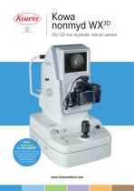
Kowa 3D nonmyd WX 2D/3D non-mydriatic retinal camera Now approved by the NDESP The Kowa nonmyd WX3D camera for retinal screening has been approved for digital photography use in Diabetic Eye Screening by the NHS Diabetic Eye Screening Programme
Open the catalog to page 1
Retinal camera Non-mydriatic Retinal camera Special function The Kowa nonmyd WX3D is a versatile retinal camera offering both stereo 3D and 2D images whilst maintaining Kowa’s values of high quality imaging and ease of use Key benefits – Quick, incredibly detailed simultaneous stereo images in just one click – Easy to use, reduced operator time – High patient throughput – Confidently assess and monitor patients suspected of having glaucoma – Accurately verifies the physiological state of the cup and disc – Proven Virtual Glaucoma Pathway solution – Efficient work flow through advanced networking...
Open the catalog to page 2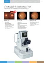
Field Angle 34° Instant & simultaneous 3D photography available in 1 shot Stereoscopic images are captured without the camera shifting Stereoscopic 3D view of the ONH (Optical Nerve Head) - Field Angle 45° - Delivers extremely detailed images with SLR - Automated 9 points fixation system allows mosaic photography covering a large retinal area and to identify peripheral pathologies - On screen guides indicate if the pupil size is within the sufficient range (above 3.5mm) for photography
Open the catalog to page 3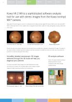
High quality 2D retinal images in one click in combination with the high resolution digital SLR camera. What’s more, the nonmyd WX3D offers a small pupil mode of 3.5mm, together with an integrated 9 point fixation system for mosaic photography covering a wider area of the retina. Examine the optic nerve quickly, easily and in far more detail than is possible from a standard 2-dimensional 2D image. This simple to use system provides a detailed quantitative display of optic disk parameters, including the vertical cup to disc ratio, neuroretinal rim area and rim to disk area along with many others....
Open the catalog to page 4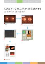
Retinal camera Non-mydriatic Retinal camera Special function Kowa VK-2 WX Analysis Software 3D analysis in 5 simple steps Photography by the Kowa nonmyd WX3D Determine the contour line for the cup & disc automatically or manually
Open the catalog to page 5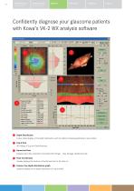
Confidently diagnose your glaucoma patients with Kowa’s VK-2 WX analysis software Depth Distribution Colour-coded display of the depth distribution, with the ability to display graphically at any position 3 Cup & Disk 3D Display of Cup and Disk flickering 3 Numerical Data Displays optic disk parameters including ‘DDLS Stage’ - Disk Damage Likelihood Scale ^4 Polar Coordinates Visually displays the location of the thinnest part of the disk rim 0 Contour line depth distribution graph Graphical display of the depth distribution of cup and disk
Open the catalog to page 6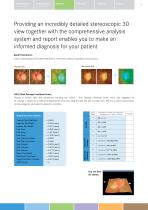
Retinal camera Non-mydriatic Retinal camera Special function Providing an incredibly detailed stereoscopic 3D view together with the comprehensive analysis system and report enables you to make an informed diagnosis for your patient Depth Distribution Colour-coded display of the depth distribution of the disk cupping and graphical cross section. Glaucoma Eye DDLS (Disk Damage Likelihood Scale) Display of various optic disk parameters including the “DDLS“ - Disk Damage Likelihood Scale; which was suggested by Dr. George L. Spaeth as a method to diagnose the optic disk using the disk size and rim/disk...
Open the catalog to page 7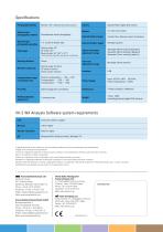
Specifications Camera Specific Nikon digital SLR camera Internal fixation target Central, Disk, Macula, mosaic 9 positions External fixation target Normal mode: 45° SP mode: 45°* Stereo mode: 34° (20° x 27°) Optical head base adjustment range Moveable 40mm forward/backward Moveable 98mm leftward/rightward Moveable 27mm vertically (electric) Chin rest adjustment range Photography Modes Normal / SP / Stereo (electrically switched) Stereoscopic photography method Simultaneous stereo photography Stereoscopic photography parallax Field angle *Some eyes may cause flare around their circumference Working...
Open the catalog to page 8All Kowa Medical Care catalogs and technical brochures
 SL-19 for veterinary
2 Pages
 SL-19
SL-192 Pages
Archived catalogs
 Non-mydriatic Fundus Camera
2 Pages
 VX-2
VX-28 Pages
 WX3D
WX3D2 Pages
 Genesis-D Brochure
2 Pages
 Nonmyd ?-DIII Brochure
2 Pages
 AP-7000 Brochure
8 Pages
 KW-2000 Brochure
1 Page
 Genesis-Df
2 Pages
 nonmyd8
1 Page
 DigiVersal
8 Pages
 VK-2-
VK-2-2 Pages
 VX-20 Brochure
8 Pages
 VX-10a Brochure
6 Pages
 FM-600
4 Pages
 2013 FM-700
4 Pages
 SL-17 Brochure
2 Pages
 SL-15L
2 Pages
 HA-2 Applanation Tonometer
2 Pages
 VK-2 WX3D
5 Pages
 DR-1a
DR-1a2 Pages
 nonmyd 7
4 Pages
 nonmyd 8
2 Pages
 nonmydAF
2 Pages
 FM-700
2 Pages
 Kowa nonmyd WX3D
2 Pages
 VX-20
VX-204 Pages


