VX-20
1 /4Pages

VX-20
1 /4Pages
Catalog excerpts

Technology for Life Science Mydriatic/ Non-Mydriatic Integrated Retinal Camera KOWA Retinal Camera Mydriatic/ Non-Mydriatic Integrated Retinal Camera Specifications Photography modes Internal fixation light Central/disc/macula/peripheral External fixation light Red/green blinking Appropriate exposure is automatically set based Exposure on the 37-steps (0.6WS to 300WS) of field angle, filter, and photography mode Camera Higher than 12 megapixel CMOS sensor Monitor 7 inch wide-screen touch LCD Forward/backward: (gross) 90mm, (micromotion) approx. 17.5mm Leftward/rightward: (gross) 140mm, (micromotion) approx. 17.5mm Adjustment range Upward/downward: 30mm Tilting: (elevation) 11°, (depression) 15° Panning: (left/right) 30° horizontally Recording medium CF memory card Interface USB : VK-2 connection Printer connection Card reader connection LAN : Image output Power saving function Yes Power supply Input : AC 120/230V 50/60Hz Options Internal fixation target Grip Auxiliary forehead rest Images in the LCD monitor are compositions. All other companies and product names stated here are trademark or registered trademark of each company. Specifications and appearances are subject to change without notice. C€ Distribution name : KOWA VX-20 Kona Company. Ltd. World Sales Headquarters 4-14, Nihonbashi-honcho 3-chome, Chuo-ku, Tokyo 103-8433 Japan Phone: +81 (3)3279-7639 Facsimile: +81 (3)3279-7541 URL: http://www. kowa. co.j p/e-1 ife/ Kowa Company, Ltd. Chofu Factory 3-1, Chofugaoka 3-chome, Chofu, Tokyo 182-0021, Japan I ec I rep I Ktfion Eluarjpe GmbH lmmermannstr. 43B, 40210 Dusseldorf, Germany Tel: +49 (0) 211-179354-18 Fax: +49 (0) 211-161952 Email: [email protected] Web site: www.kowamedical.com Kowa American Corporation Medical Devices Division 20001 South Vermont Avenue, Torrance, CA 90502, U.S.A Tel: +1(310)-327-1913 FAX: +1(310)-327-4177 E-mail: [email protected] URL: http://kowa-usa.com/
Open the catalog to page 1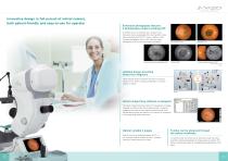
Retinal Camera both patient-friendly and easy-to-use for operator Enhanced photography features 5 photography modes including FAF* In addition to the non-mydriatic color, mydriatic color, fluorescein fundus angiography (FA), Red-free (RF) modes, fundus autofluorescence (FAF*) mode is added to allow 5-modes photography. Also, the SP mode that allows photography of 03.5 mm of small pupil is equipped. Updated design providing stress-free diagnosis 75mm reduced height of examined eye from our previous model ("VX-10 " series), allowing photography in a relaxed posture for patient. Instant image filing...
Open the catalog to page 2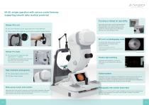
VX-20, simple operation with various useful features, supporting smooth daily medical practices!Focusing on design for operability Design:Chin rest Only usable switch buttons are illuminated according to each photography mode, enabling smooth and quick ® Eye level indication can be easily seen even in the darkroom. ® Stable chassis that makes it easy to assist in patient's eyelid opening. photography even in the darkroom. Moving chin rest up/down and switching field of view are electrically performed, that can be operated at hand. SP (Small Pupil) photography mode In mydriatic mode, either 03.5mm...
Open the catalog to page 3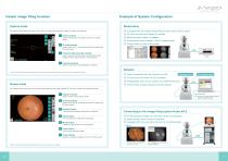
Retinal Camera Example of System Configuration Capture mode The capture mode allows the user to Input IDs and display the alignment Images or Images just captured. Used to switch from the capture mode to the viewer mode. Q ID input button Used to Input the ID. Pressing this button to display the ID Input screen. Q Fixation light selection button Used to select the fixation light. Pressing this button displays fixation light buttons to allow selection. 0 Equipped with the instant image filing function using a touch panel. 0 ID input from the card reader is possible. 0 Multiple-image display (4...
Open the catalog to page 4All Kowa Medical Care catalogs and technical brochures
 SL-19 for veterinary
2 Pages
 SL-19
SL-192 Pages
Archived catalogs
 Non-mydriatic Fundus Camera
2 Pages
 VX-2
VX-28 Pages
 WX3D
WX3D2 Pages
 Genesis-D Brochure
2 Pages
 Nonmyd ?-DIII Brochure
2 Pages
 AP-7000 Brochure
8 Pages
 KW-2000 Brochure
1 Page
 Genesis-Df
2 Pages
 nonmyd8
1 Page
 DigiVersal
8 Pages
 VK-2-
VK-2-2 Pages
 VX-20 Brochure
8 Pages
 VX-10a Brochure
6 Pages
 FM-600
4 Pages
 2013 FM-700
4 Pages
 SL-17 Brochure
2 Pages
 SL-15L
2 Pages
 HA-2 Applanation Tonometer
2 Pages
 VK-2 WX3D
5 Pages
 DR-1a
DR-1a2 Pages
 nonmyd 7
4 Pages
 Nonmyd WX3D
8 Pages
 nonmyd 8
2 Pages
 nonmydAF
2 Pages
 FM-700
2 Pages
 Kowa nonmyd WX3D
2 Pages


