Group: KOWA Group

Catalog excerpts
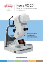
Combined mydriatic & non-mydriatic retinal camera 7 in 1 hybrid retinal imaging system Versatile 7 in 1retinal imaging system (FAF, FA, RF, colour, anterior, mydriatic and non-mydriatic and 9 point internal fixation targets for mosaic imaging)
Open the catalog to page 1
The Kowa VX-20 is a unique, hybrid combined retinal camera that offers versatile, high quality, retinal imaging in one easy to use system Due to its outstanding features, the Kowa VX-20 remains the first choice for clinicians - Superior quality retinal imaging - Ergonomic design for enhanced patient comfort and operator experience - Simple, easy to use system - Fast image capture in FA mode - Saves time with direct printer & network connections - Saves space with modern, compact design - Narrow camera design allows the operator better view of the patient - Efficient work flow through...
Open the catalog to page 2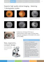
Retinal camera Non-mydriatic Retinal camera Special function Superior high quality retinal imaging – featuring 7 photography modes Mydriatic colour Non-mydriatic colour Fluorescein fundus angiography (FA) (the actual captured images include the timer) Small pupil mode (the actual captured images include the timer) Furthermore, the Kowa VX-20 has an autofluorescence (FAF) mode to provide further diagnostic investigation of the health of the retina such as ARMD. Anterior New, ergonomic, compact design The new innovative design of the VX-20 ensures both patient comfort and ease of use for the...
Open the catalog to page 3
Retinal camera Non-mydriatic Retinal camera Special function Easy to use, efficient retinal camera supporting your daily practice within the ophthalmic environment Relaxed, comfortable diagnosis The new position for your patients’ eye is 75mm lower than Kowa’s previous model enabling a more comfortable, relaxed position. Kowa VX-10a (previous model) Easy operation - even in darkrooms The stable chassis offers easier patient alignment whilst the LED eye level mark makes testing easier in darkrooms.
Open the catalog to page 4
Retinal camera Non-mydriatic Retinal camera Special function Larger range of peripheral imaging The downward tilt angle has been increased to 11° increasing the possible range of peripheral imaging. Easy patient observation The Kowa VX-20 includes a 7 inch wide, LCD colour touch screen that is simple to use and is ideally positioned to ensure easy patient observation. Simple 1-handed operation The control panel for the Kowa VX-20 has been designed in order to improve the operator experience and keep everything within easy reach for the clinician. Features – Operational buttons positioned...
Open the catalog to page 5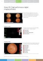
Kowa VK-2 high performance digital imaging software The Kowa VK-2 digital imaging software easily captures and stores retinal photographs taken from the Kowa VX-20 retinal camera, producing high quality 2D retinal images in just one click in combination with the high resolution digital SLR camera. The capture mode allows the user to input IDs and display the alignment images or images just captured. [J Viewer Button ^ ID input button [3 Fixation light selection button r3 Custom buttons The viewer mode allows the user to browse the images saved in the selected ID, and print or delete the...
Open the catalog to page 6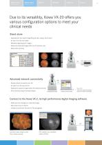
Retinal camera Non-mydriatic Retinal camera Special function Due to its versatility, Kowa VX-20 offers you various configuration options to meet your clinical needs Stand alone – Equipped with the instant image filing function using a touch panel – ID input from the card reader – Multiple-image display (4 images) – Easily save captured images within the CF memory card – Allows direct printing Advanced network connectivity – Simple intranet connection via LAN – ID input from the Kowa VX-20 – Easily store captured images within the network terminal – Direct printing using the preview display...
Open the catalog to page 7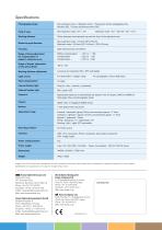
Specifications Photography modes Non-mydriatic colour / Mydriatic colour / Fluorescein fundus angiography (FA) Red-free (RF) / Fundus autofluorescence (FAF) Working distance 39mm (between the examined eye and the front of the objective lens) Minimum pupil diameter Split luminous bars coincidence Range of focus adjustment for compensation of patient’s refractive error Without compensation : -12D to + 13D Compensation – : - 32D to - 10D Compensation + : +10D to +35D Range of diopter adjustment of the optical finder Working distance adjustment Luminous dot indication (ON / OFF switchable)...
Open the catalog to page 8All Kowa Optimed catalogs and technical brochures
-
 VX-20
VX-204 Pages
-
 Kowa nonmyd WX3D
2 Pages
-
 FM-700
2 Pages
-
 nonmydAF
2 Pages
-
 nonmyd 8
2 Pages
-
 Nonmyd WX3D
8 Pages
-
 nonmyd 7
4 Pages
-
 DR-1a
DR-1a2 Pages
-
 VK-2 WX3D
5 Pages
-
 HA-2 Applanation Tonometer
2 Pages
-
 SL-15L
2 Pages
-
 SL-17 Brochure
2 Pages
-
 FM-700
4 Pages
-
 FM-600
4 Pages
-
 VX-10a Brochure
6 Pages
-
 VK-2-
VK-2-2 Pages
-
 DigiVersal
8 Pages
-
 nonmyd8
1 Pages
-
 Genesis-Df
2 Pages
-
 KW-2000 Brochure
1 Pages
-
 AP-7000 Brochure
8 Pages
-
 Nonmyd ?-DIII Brochure
2 Pages
-
 Genesis-D Brochure
2 Pages
-
 WX3D
WX3D2 Pages
-
 VX-2
VX-28 Pages
-
 Non-mydriatic Fundus Camera
2 Pages


