- Catalogs
- KUBTEC Medical Imaging
- The MOZART® Specimen Tomosynthesis Imaging System
The MOZART® Specimen Tomosynthesis Imaging System
1 /8Pages

The MOZART® Specimen Tomosynthesis Imaging System
1 /8Pages
Catalog excerpts

Margin Management Without All the False Positives Intraoperative Specimen Tomosynthesis MEDICAL IMAGING
Open the catalog to page 1
“I believe that 3D tomosynthesis specimen X-ray is more accurate. It helps us beyond the other generation of two-dimensional imaging. Good for the patient because if we can be more accurate, of course it reduces the re-excision rate.” “We want the patient to have the best possible results, a combination of least times having to go back for cancer or re-excision, but taking out the least amount of tissue to preserve the best cosmesis. So I think having 3D specimen X-rays is letting us do both those things." Peter Blumencranz, MD, FACS Medical Director The Comprehensive Breast Care Center of Tampa...
Open the catalog to page 2
“For my practice, and many breast cancer centers of excellence around the world... Using 3D specimen tomosynthesis during surgery has helped the best surgeons reduce their re-excision rates even more.” - U.S. News & World Report October 30, 2019 Sheldon M. Feldman, MD, FACS Chief of Breast Surgery and Breast Surgical Oncology and Director of Breast Cancer Services at Montefiore Health System, New York, NY "3D tomography has radically streamlined breast cancer surgery by allowing surgeons to better visualize the breast and affected area, even through dense breast tissue, in the operating room.”...
Open the catalog to page 3
The MOZART® System is more accurate at identifying positive margins. igital Breast Tomosynthesis is rapidly gaining acceptance as the next gold standard for specimen mammography. Unlike its traditional 2D X-ray counterpart, tomosynthesis allows the physician to see the breast specimen in 3 dimensions which improves accuracy and reduces false positives. A depiction of a lumpectomy that has a lesion on a peripheral margin. In a traditional 2D specimen X-ray, the three dimensional anatomy is compressed into a single planar view. All vertical perspective is lost. In this example the positive margin...
Open the catalog to page 4
A depiction of a lumpectomy that has a lesion on a peripheral margin. Benefits of 3D Tomosynthesis Specimen tomosynthesis enables analysis of the specimen in 1 millimeter digital slices. Each slice anatomically has its own margin, and can be viewed independently of all the other slices. The view of each slice is unobscured by dense tissue above or below. Specimen tomosynthesis creates 1mm digital slices, each independent of the others. The peripheral lesion is on the 4mm slice. The surgeon is able to analyze the location and the extent of the lesion, and involvement of the peripheral, anterior...
Open the catalog to page 5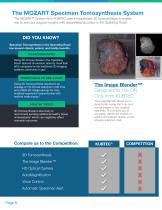
DID YOU KNOW? Specimen Tomosynthesis in the Operating Room has several clinical, patient, and facility benefits. RE-EXCISION RATES Using 3D tomosynthesis in the Operating Room reduces re-excision rates by more than 50% compared to the traditional 2D imaging systems commonly in use.1 REDUCTION IN OR TIME & COST Using 3D Tomosynthesis resulted in an average of 7.6-minute reduction in OR time and a $284.62 charge savings for wire-localized segmental mastectomies with sentinel node biopsy.2 HEALTHY TISSUE 3D Tomosynthesis is less likely to recommend excising additional healthy tissue unnecessarily3...
Open the catalog to page 6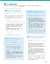
Clinical Studies Specimen Tomosynthesis is a breakthrough technology for intraoperative quality of care. Clinical research suggests that The MOZART® System can help reduce re-excision rates and improve cosmesis for your patients. Study: Differences in Re-excision rates for Breast-Conserving Surgery Using Intraoperative 2D Versus 3D Tomosynthesis Specimen Radiograph Natalia Partain, MD, Carissia Calvo, MD, Ali Mokdad, MD, Andrea Colton, MD, Katherine Pouns, MD, Edward Clifford, MD, Deborah Farr, MD, James Huth, MD, Rachel Wooldridge, MD, A. Marilyn Leitch, MD. Ann Surg Oncol 2020 : Published online...
Open the catalog to page 7
3D Specimen Imaging Call 1.203.364.8544 to set up a demo for your operating room. The MOZART® Supra® Specimen Tomosynthesis System Specimen Tomosynthesis System Calculate the Savings for Your Operating Room. This is intended to be an example of potential savings for the Operating Room. All figures will be adjusted based on your needs. 1. Colton A, Calvo C, Mokdad A, Pouns K, Clifford E, Farr D, Huth J, Wooldridge R, Leitch M, Partain N. Differences in Re-excision Rates for Breast Conserving Surgery Using Intraoperative 2D vs. 3D Tomosynthesis Specimen Radiograph. Poster presented at: American...
Open the catalog to page 8All KUBTEC Medical Imaging catalogs and technical brochures
 The XPERT® 80/80-L Brochure
8 Pages
 The XPERT® 40 Brochure
2 Pages
 The XPERT® 20 Brochure
2 Pages