
- Catalogs
- KUBTEC Medical Imaging
- The XPERT® 80/80-L Brochure

- Company
- Products
- Catalogs
- News & Trends
- Exhibitions
The XPERT® 80/80-L Brochure

The XPERT® 80/80-L Brochure
KUBTEC Imaging Systems are designed to enhance patient care in anatomic pathology by improving efficiency and reducing turnaround times in the Gross Room. These systems, including the XPERT® 80/80-L and MOZART® Supra®, allow for on-site imaging, eliminating the need to transport specimens to radiology.
Key Features and Benefits
- Efficiency: Single-click image capture and rapid viewing reduce time spent on locating biopsy clips and other specimen details.
- Safety: Fully shielded systems ensure operator safety, and the ability to see lesions or clips before cutting reduces the risk of cutting through hardware.
- Accuracy: On-site imaging increases the accuracy of cuts and reduces the risk of lost specimens.
- Cost Reduction: Decreased workload on histology staff and reduced supply utilization lead to long-term cost savings.
Applications
- Breast Imaging: Instantly locate biopsy clips.
- Organ Imaging: Applicable to liver, lung, spleen, kidney, and more.
- Bone Imaging: Identify hardware before using a bone saw.
- Fetal Demise and Autopsy: Measure skeletal deformities and locate stents in heart specimens.
Advanced Imaging Technology
- 3D Tomosynthesis: Provides detailed 3D views of specimens, allowing for precise assessment of clips, calcifications, and lesions.
- Image Blender™: Overlays X-ray and optical images for accurate cuts.
- Automatic Specimen Alert: Alerts if a specimen is not removed after imaging.
- Voice Control and AutoMagnification: Enhance user interaction and image quality.
System Specifications
- Detector Sizes: Vary across models, offering options for different imaging needs.
- Spatial Resolution: Up to 20 lp/mm in contact mode for high-resolution imaging.
Conclusion
KUBTEC Imaging Systems provide significant improvements in pathology workflow, accuracy, and safety, making them a valuable addition to any pathology department. The systems are designed to be user-friendly, requiring no specialized X-ray knowledge, and offer a range of features that enhance the imaging process.
Catalog excerpts

Improving Patient Care in Anatomic Pathology. KUBTEC Imaging Systems for the Gross Room KUBTEC MEDICAL IMAGING
Open the catalog to page 1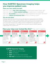
How KUBTEC Specimen Imaging helps you improve patient care. Have you ever asked yourself: How can I spend less time searching for that elusive breast biopsy clip? How can I save the time wasted walking my specimens to radiology? How can I be certain that all the bone screw fragments have been removed? How can I reduce turnaround time without increasing costs? You are not alone. Pathology departments around the world are using Specimen Imaging Systems from KUBTEC right there in the Gross Room to improve efficiency, reduce turnaround times and improve safety. The XPERT® 80/80-L and MOZART® Supra®...
Open the catalog to page 2
KUBTEC Imaging: Breast, Organs, Bone & More • • • • • Breast: Instantly locate biopsy clips. Organs: Liver, lung, spleen, kidney, adrenals, pancreas, ovaries and uterus. Bone: Identify and locate hardware before you use the bone saw. Fetal Demise: View and measure skeletal deformities. Autopsy: Locate stents in coronary vessels in heart specimens. KUBTEC X-ray imaging in the gross room clearly shows surgical staples in these images of a colon. The integrated HD camera automatically gives optical images. Fig A shows a loop of previously anastomosed bowel and mesenteric fat showing the anastomotic...
Open the catalog to page 3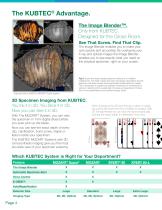
The Image Blender™. Only from KUBTEC. Designed for the Gross Room. See That Screw. Find That Clip. The Image Blender enables you to make your cuts quickly and accurately. By overlaying your X-ray and optical images the Image Blender enables you to see exactly what you need on the physical specimen, right on your screen. Fig C shows the deep margin (posterior surface) of a modified mastectomy. The deep margin has been previously inked black and the specimen has been serially sectioned (bread loafed) lateral to medial. This is important to assess total disease involvement 3 dimensions as well as...
Open the catalog to page 4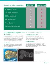
KUBTEC® COMPETITION Automatic Specimen Alert ✓ HD Optical View Only KUBTEC Systems include an integrated HD optical camera which automatically takes an optical image of your specimen at the exact time it creates the X-ray image. Essential for ensuring specimen orientation and accuracy. Voice Control Use voice commands for complete control over image viewing and navigation without touching the keyboard. Automatic Specimen Alert Automatic Specimen Alert on all KUBTEC Systems uses the integrated optical camera to alert you if the specimen is not removed after the X-ray image is taken. AutoMagnification...
Open the catalog to page 5
Let Us Help You. Trying to change the way your gross room runs is a challenge. Let us help. With our Pathology Savings Calculator, we can show you and your team the savings that a KUBTEC specimen imaging system can provide to your lab. From reducing block submission and recuts to time savings, we can provide one-on-one assistance to support your business case. “Our Surgical Team and Pathology Director were impressed by the improvements in turnaround time. The image quality is excellent and KUBTEC has so many more features that truly make a difference in how we do our job - it was a no-brainer”....
Open the catalog to page 6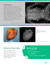
Fetal Demise X-Ray imaging is often recommended or mandated in cases of fetal demise. The KUBTEC XPERT 80-L has an imaging area large enough to image a complete full-term fetus. The high-resolution system provides rapid, highly detailed images to measure gestational assessment, malformations, skeletal dysplasia. What’s more the system allows you to take essential measurements directly on the image, and the optical camera and Image Blender™ enable you to see internal structures while viewing an optical image of the specimen. Captured with a KUBTEC XPERT 80-L System Fig E Human heart X-ray with...
Open the catalog to page 7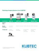
Specifications XPERT® 80 XPERT® 80-L MOZART® MOZART® Supra® Detector 10” x 12” (23 x 29 cm) 17” x 17” (43 x 43 cm) 5” x 6” (12 x 15cm) 10” x 12” (23 x 29 cm) Sizes Spatial Resolution up to 10 Ip/mm, 20 Ip/mm, contact mode 10 Ip/mm, contact mode 10 Ip/mm, contact mode contact mode Imaging Type 2D Imaging OpticaI Imaging 2D Imaging Optical Imaging 3D Tomosynthesis 2D Imaging Optical Imaging 3D Tomosynthesis 2D Imaging Optical Imaging The Image Blender is a trademark, and Kubtec, the Kubtec logo, XPERT, and MOZART are registered trademarks of KUB Technologies, Inc. M1291A-0620 KUBTECMEDICAL IMAGING...
Open the catalog to page 8All KUBTEC Medical Imaging catalogs and technical brochures
 The XPERT® 40 Brochure
2 Pages
 The XPERT® 20 Brochure
2 Pages
