- Catalogs
- LargeV Instrument
- Ultra3D
- Company
- Products
- Catalogs
- News & Trends
- Exhibitions
Ultra3D
1 /12Pages

Ultra3D
1 /12Pages
Catalog excerpts
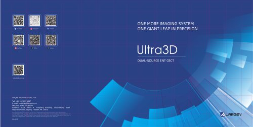
ONE MORE IMAGING SYSTEM ONE GIANT LEAP IN PRECISIONUltra3DDUAL-SOURCE ENT CBCT LargeV Instrument Corp., Ltd. Tel: +86-10-5083 6847 E-mail: [email protected] Website: www.largev.com Address: 800B, Block A, Tongfang Building, Shuangqing Road, Haidian District, Beijing, 100084, RR. China This document is provided to you for your information and discussion only. Any information including functions, images or dimensions, may be changed or summarised and is expressed as of the date of writing. The information may change without notice and LargeV Instrument Corp., Ltd. Is under no obligation to ensure that such updates are brought to your attention. (20230616)
Open the catalog to page 1
Ultra3D THE MICRON-SCALE IMAGING \NORLD
Open the catalog to page 2
Using a dual-source generator and dual-detector design, Ultra3D contributes to a dual-system with wide-angle imaging and microscopic imaging that can provide micron-scale imaging solutions for ENT and dental clinical diagnosis. Ultra3D captures a large area with 2D image after will be select small area for 3D shooting ,it will automatically direct the patient to the correct position with easy manipulation MICROSCOPIC IMAGING SYSTEM
Open the catalog to page 3
EXTRAODINARY IMAGE QUALITY It provides a ten-micron class definition, body construction with 50um can be detect easily, for in-depth diagnostic. STARRY SKY DETECTOR Equipped with high resolution, high sensitivity, high image acquisition rate, and a low power consumption core. 0.25 SMALL FOCAL SPOT X-RAY TUBE Core technology for improving resolution and SNR. THREE SUPPORTIVE ALGORITHMS HD Reconstruction Scatter Correction Metal Artifact Reduction 0.25 SMALL FOCAL SPOT X-RAY TUBE LOW DOSE CONTROL It fully explores the perfect balance between image quality and radiation dose. Low dose mode provides...
Open the catalog to page 4
HUMAN-MACHINE INTERACTION FRIENDLY MORE PRECISE MORE CONVENIENT Comfortable Table The scanning table follows ergonomics to achieve smooth movement in six directions: up and down, left and right, and front and back, with 0.1 mm movement control accuracy. To reduce motion artifacts, the patients remain stable and comfortable during the whole procedure. Optimaize positioning workflows with dramatically simplified laser projection system if the patient is positioned as accurate as possible. Interaction System for Communication Doctors can communicate with patients in real time. The console can control...
Open the catalog to page 5
3D Fine Reconstruction Minimum voxel size to 0.05 mm. Neural Tube Automatic Labeling Label the neural tube automatically in the CT image, providing great convenience for diagnosis. Airway Measurements Automatically calculate the volume and the narrowest area of the airway in the form of chromatographic visualization. Quick Film Printing It can quickly set the layout and spread the film quickly. Also,it can measure the film and adjust the window value and other functions. Medical Images Comparison By loading multiple images, it is possible to perform single-slice comparison medical imaging. It...
Open the catalog to page 6
The CT image resolution is up to ten-micron class definition with Ultra3D. The internal structure of the ear can be visualized by a HD image, vastly increasing the detection capability. The microscopic imaging system also provides high-quality images for endodontic, periodontal and TMJ. The wide-angle imaging system can fulfill orthodontics, maxillofacial surgery, and clinical implant diagnosis.
Open the catalog to page 7
CLINICAL APPLICATIONS IN OTOLOGY Pulsatile Tinnitus Ultra3D clearly shows the Ultra3D shows dural arteriovenous fistula vascular branch, diverticulum missing. Artificial Auditory Ossicle Implantation Measured by Ultra3D: Artificial auditory ossicle on the side of the vestibule is 1.1 mm deep, the shortest distance to the intine is 1.9 mm, the distance to the edge of the vestibular window is between 1.1 mm and 1.2 mm, and the long angle of the incus is 101°. Cochlear Implant Surgery Pre-operation CTA+CTV: Sigmoid sulcus missing. Ultra3D shows the fine repayment of the sigmoid sulcus.
Open the catalog to page 8
Cariology, Endodontics and TMJ Examination The microscopic imaging system is equipped with an unique small FOV reconstruction algorithm that can achieve high-definition reconstruction with a minimum spatial resolution of 50 urn. It can be used as an imaging tool for endodontic diseases, dentition restoration, and joint diseases, which is of great significance for diagnosing and analyzing fine anatomical structures in diseases. For example, observing small branches of the facial nerve canal, diagnosing early lesions such as fissures and fractures, and diagnosing TMJ. The cortical wear and surface...
Open the catalog to page 9
PRODUCT PARAMETERS X-ray Generator 2 Nominal Focal Spot Size Adjacent Tube Voltage Increment 5kV Tube Voltage Adjacent Tube Voltage Increment 5kV Adjacent Tube Current Increment 0.5mA| Tube Current Adjacent Tube Current Increment 0.5mA Maximum Power II Flat Panel Detector 1 Imaging System Field of View (Diameter x Height) Spatial Resolution (Ip/mm) Wide-angle Imaging System 23x19 16x13 16x6 Microscopic Imaging System Flat Panel Detector 2 Cesium Iodide + CMOS Effective Imaging Area Pixel Size Acquisition Matrix
Open the catalog to page 10
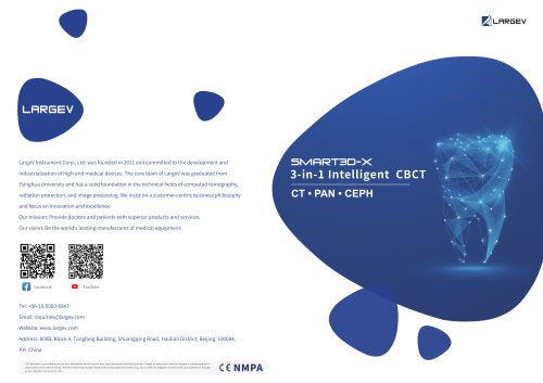
LargeV Instrument Corp., Ltd. was founded in 2011 and committed to the development and industrialization of high-end medical devices. The core team of LargeV was graduated from Tsinghua University and has a solid foundation in the technical fields of computed tomography, radiation protection, and image processing. We insist on a customer-centric business philosophy and focus on innovation and excellence. Our mission: Provide doctors and patients with superior products and services. Our vision: Be the world's leading manufacturer of medical equipment.
Open the catalog to page 11
2013 Passed TuV ISO 13485 quality management system certification and CE certification. Achieved the title of "National High-tech Enterprise." Awarded the first level prize of "Technological Invention" by the Chinese Society for Stereology Science and Technology. The first Chinese CBCT debuted at the International Dental Show (IDS) in Cologne, Germany. The Multifunction Dental CBCT Smart3D was granted certification from NMPA. HiRes3D-Plus and HiRes3D-Max, professional dental CBCT models with super-large FOV were certified by NMPA. Awarded with "Edgy Technology Enterprise" by Beijing Pharmaceutical...
Open the catalog to page 12
Archived catalogs

- Analysis software
- Visualization software
- Radiology software
- Tablet computer software
- Tablet PC software
- Diagnostic software
- Planning software
- Hospital software
- Dental software
- Design software
- Dental radiography system
- Treatment software
- Data management software
- Measurement software
- Digital dental radiography system
- Simulation software
- Sharing software
- Server software
- CT software
- Evaluation software