
- Catalogs
- Leica Biosystems
- The Leeds Guide to Digital Pathology Volume 2


- Company
- Products
- Catalogs
- News & Trends
- Exhibitions
The Leeds Guide to Digital Pathology Volume 2

The Leeds Guide to Digital Pathology Volume 2
The document introduces the second volume of "The Leeds Guide to Digital Pathology," highlighting the advancements in digital pathology since 2018, particularly during the COVID-19 pandemic. It emphasizes the development of a national digital pathology network and its role in maintaining high-quality diagnostic services.
This section stresses the importance of maintaining quality in digital pathology labs, focusing on ISO-15189 accreditation. Key aspects include planning, change control, health and safety assessments, and ensuring image comparability and reproducibility. Regular scanner maintenance and calibration are crucial for quality assurance.
The clinical applications of digital pathology are explored, highlighting its integration into diagnostic workflows and the benefits of increased efficiency and accuracy.
The potential for pathologists to report from home is discussed, facilitated by digital pathology. Necessary infrastructure and protocols to support this practice are outlined.
This section outlines the deployment of digital pathology across multiple sites, forming national networks to enhance collaboration and standardization in pathology services.
The role of AI in digital pathology is examined, focusing on its potential to improve diagnostic accuracy and efficiency, and the importance of integrating AI tools into pathology workflows.
The importance of involving patients and the public in the development and implementation of digital pathology services is emphasized to ensure transparency and trust.
St James University Hospital in Leeds has integrated digital pathology into its workflows, with all histology slides scanned and accessible on diagnostic workstations. The transition began with breast and neuropathology and expanded to other specialties, underscoring the value of digital slides for remote work and collaboration during the pandemic.
A training and validation program for digital pathology was developed, ensuring pathologists can confidently make digital diagnoses, focusing on individual comfort and skill development.
The deployment of a national vendor-neutral archive is recommended to centralize storage and reduce costs, facilitating case sharing and standardizing workflows. Emphasizing standards and interoperability is crucial for integrating various slide scanners and software systems.
Effective communication is crucial to address public mistrust and ensure patients understand the relevance and benefits of digital pathology. Patients often perceive digital data as less secure than paper records.
Digital pathology has transformed diagnostic processes, offering efficiency, flexibility, and enhanced collaboration. The future includes integrating complex image analysis and AI to further improve diagnostic quality and efficiency. The document concludes with contact information for the authors and acknowledges the support from various organizations and partnerships in advancing digital pathology research and deployment.
Catalog excerpts

The Leeds Guide to Digital Pathology Volume 2 Building a National Digital Pathology Network National Pathology Imaging Co-operative
Open the catalog to page 1
INTRODUCTION P3 CHAPTER 1 – MAINTAINING QUALITY IN THE LABORATORY P5 CHAPTER 2 – DIAGNOSTIC DIGITAL PATHOLOGY: CLINICAL USE P13 CHAPTER 3 – HOME REPORTING P21 CHAPTER 4 – MULTISITE DIGITAL PATHOLOGY DEPLOYMENT AND NATIONAL NETWORKS P29 CHAPTER 5 – ARTIFICIAL INTELLIGENCE P37 CHAPTER 6 – PATIENT AND PUBLIC INVOLVEMENT AND ENGAGEMENT P43 FINAL THOUGHTS P47 FURTHER RESO
Open the catalog to page 2
INTRODUCTION It’s hard to believe how the world has transformed in the three years since the team first published “The Leeds Guide to Digital Pathology” in 2018. The COVID-19 pandemic forced everyone, including the pathology community to rise to new challenges. The flexibility and resilience offered by digital pathology have never seemed more important if we want to future-proof our diagnostic services and ensure we can continue to deliver a high-quality histopathology service in a timely manner. At Leeds, we were fortunate to have completed our initial scanner deployment of Aperio AT2 scanners,...
Open the catalog to page 4
The foundation of quality digital diagnosis lies in the laboratory, where biomedical staff work exceptionally hard, often under challenging time constraints.
Open the catalog to page 5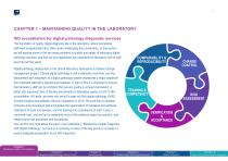
CHAPTER 1 – MAINTAINING QUALITY IN THE LABORATORY ISO accreditation for digital pathology diagnostic services The foundation of quality digital diagnosis lies in the laboratory, where biomedical staff work exceptionally hard, often under challenging time constraints. In this section, we will explore some of the key areas pertinent to quality and safety of laboratory digital pathology services, and find out how digitisation has impacted the laboratory and its staff over the last five years. Digital pathology deployment in the clinical laboratory represents a massive change management project....
Open the catalog to page 6
Key tips 1. e prepared! Careful planning in the earliest stages of your deployment B can help lay the groundwork for successful inspection and accreditation. Identify key personnel who will be instrumental in delivering core aspects of the accreditation procedure, and compiling necessary evidence. 2. evelop and adhere to a full change control procedure. This should D include evidence surrounding the digital deployment proposal, health and safety assessments, validation and verification of equipment, external and internal quality assurance processes, IT user acceptance testing, standard operating...
Open the catalog to page 7
Quality assurance Daily (Recommended) • Restart the scanner and console (PC) if present • Clean the touchscreen • Clean the scanner cover and light source • Clean the slide trays and carousel/loader • Sections should be free of folds, creases and bubbles. • issue should be placed centrally, within the scannable margins of T the glass slide. • Clean the objective and Koehler • Clear the slide stage tray • Clean the fan filter When Required • Light Source bulb replacement • Schedule annual maintenance visit by vendor The key steps to quality assurance of scanned slides are relatively simple, and...
Open the catalog to page 8
Quality control in the laboratory Laboratory quality control is an important step in the histopathology process as it checks that digital slides being signed out of the laboratory are of adequate diagnostic quality. At digital QC the lab are checking for: • overall image quality • gross or focal loss of focus and the reason for this • are there areas of the image out of focus, and the reason Using digital images for the full laboratory quality control pathways Some laboratories check their glass slides for section and staining quality using standard light microscopy before they are sent to pathologists....
Open the catalog to page 9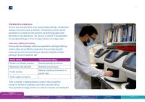
Interlaboratory comparisons As more and more laboratories move towards digital pathology, interlaboratory schemes will become easier to establish. Interlaboratory schemes enable laboratories to compare how their scanners are performing against other laboratories in the network/area. This ensures an element of standardisation across digital pathology in terms of image production and image quality. Laboratory staffing and training Ensuring staff are adequately trained and supported to use digital pathology systems safely and confidently is paramount. A two-pronged approach incorporating vendor...
Open the catalog to page 10
PERSONAL PERSPECTIVE Sarah Caton
Open the catalog to page 11
We’ve seen a lot of change in the laboratory since 2015, evolving from a single scanner installation to running a full clinical service, scanning 100% of our glass slides. We’ve implemented next generation scanners – four Aperio GT 450 DX scanners, which have changed the way we work. We have observed an 85% improvement in scanning efficiency compared with our previous scanners, and we have been able to incorporate digital pathology into other workflows including our autosectioner. One thing I would emphasize, is how important it is to try and keep everyone in the laboratory engaged and involved...
Open the catalog to page 12
“Digital pathology has improved my turn around time for diagnosis by at least 30 seconds for each slide. It’s very easy and quick to navigate between areas in the same slide and between slides in a case, saving me time and effort navigating through large and complex cases. Dr Azzam Ismail Consultant Neuropathologist Leeds Teaching Hospitals NHS Tr
Open the catalog to page 13
CHAPTER 2 – DIAGNOSTIC DIGITAL PATHOLOGY: CLINICAL USE St James University Hospital, Leeds, UK, is a major NHS cancer centre serving a population of 3 million patients, with a fully subspecialised diagnostic histopathology department. We’ve been integrating digital pathology into our standard clinical workflows over the past 5 years. 100% of histology slides are scanned in the laboratory, and accessible by our consultant and trainee pathologists on medical grade diagnostic workstations. Our first subspecialties to convert to digital microscopy for primary diagnosis were breast and neuropathology,...
Open the catalog to page 14All Leica Biosystems catalogs and technical brochures
 APERIO VERSA
4 Pages
 Aperio CS2
3 Pages
 Aperio WebViewer DX User’s Guide
84 Pages
 Aperio LIS Connectivity Overview
70 Pages
 Aperio eSlide Manager
100 Pages
 APERIO CLINICAL SOLUTION
7 Pages
 APERIO I M AG E A N A LYS I S
20 Pages
 Leica ASP6025 S
4 Pages
 Leica VibratomeTM Series
8 Pages
 Aperio GT 450 DX
5 Pages
 BOND RXm
4 Pages
 Leica St4020 Linear Stainer
2 Pages
 HistoCore SPECTRA ST Stainer
4 Pages
 Leica CM1520 Cryostat
2 Pages
 Leica SM2010 R
4 Pages
 Vibrating Blade Microtomes
8 Pages
 HistoCore PEARL
4 Pages
 CEREBRO
8 Pages
 HistoCore PELORIS 3
3 Pages
 Leica CM3050 S
4 Pages
 2019 HistoCore Microtomes
8 Pages
 Leica ST5010 Autostainer X
8 Pages
 Leica CM1860/CM1860 UV
5 Pages
 Aperio eSlide Manager
4 Pages
 Leica CM1950
12 Pages
 HistoCore BIOCUT
2 Pages
 HistoCore Microtomes
4 Pages
 Stereotaxic Solutions
7 Pages
 HistoCore PERMA S
4 Pages
 Leica CM3600 XP
2 Pages
 Leica RM2125 RTS
2 Pages
 Leica TP1020
4 Pages
 Leica ASP300 S
8 Pages
 Leica IP C
12 Pages
 Cognitive Cxi
2 Pages


- Leica solution reagent
- Molecular biology reagent
- Leica research reagent
- Leica laboratory reagent
- Analysis software
- Leica diagnostic reagent
- Protein reagent
- Enzyme reagent
- Leica histology reagent
- Leica stain reagent
- Leica cytology reagent
- Leica sample preparation system
- Wardrobe with drawer
- Leica automatic sample preparation system
- Leica buffer solution reagent
- Leica immunoanalysis reagent
- Leica medium reagent
- Leica antibody
- Reporting software
- Laboratory software