 Website:
Leica Microsystems
Group: Danaher
Catalog excerpts

DM1000 – DM3000 ERGONOMIC SYSTEM MICROSCOPES MYcroscopy: Designed to adapt to Your individual Daily Routine The DM1000 is ideally suited for screening clinical laboratory applications, such as histopathology, cytology, hematology, and microbiology. The DM2000 is designed for more complex routine pathology and cytology laboratory applications. Height-adjustable focus knobs allow hands and forearms to rest comfortably on the bench, independent from individual hand sizes. One hand operation of focus knob and stage drive to speed up your operation, and to free one hand for other tasks. 0-35°...
Open the catalog to page 2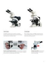
The DM2500 is especially suited for applications in pathology or biomedical research that frequently require special contrast methods, such as fluorescence or interference contrast. With its intelligent automation, the DM3000 is designed primarily for cytology and pathology laboratories in which fast work is the order of the day without sacrificing user comfort. Experience optimal ergonomic operation of a microscope thanks to symmetrical layout of coaxial drive and focus knobs. Shoulders are level, the spine is straight and arms are resting at a comfortable angle without stretching....
Open the catalog to page 3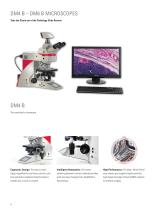
DM4 B – DM6 B MICROSCOPES Take the Strain out of the Pathology Slide Review Ergonomic Design: The easy to reach stage, magnification and focus controls, plus fully automatic condenser head movement, enables you to work in comfort. Intelligent Automation: One-button switching between contrast methods provides quick and easy changing from brightfield to fluorescence. High Performance: The large, 19mm field of view camera port supports highly sensitive, high speed and larger format sCMOS cameras for brilliant imaging
Open the catalog to page 4
DM6 B Powerful clinical upright microscope solution Fluorescence Imaging: The unique and patented Fluorescence Intensity Manager (FIM) facilitates easy and reproducible regulation of the excitation light, which helps protect your sample from photo bleaching. Rapid Measurements: The 1.25x objective coupled with a wide field of view enables users to view large specimens in a single overview.
Open the catalog to page 5
Key facts: > Convenience with LED transmitted light illumination for constant color temperature > DM2000-3000 feature a sophisticated focus mechanism -2-gear or optional 3-gear focusing, with torque adjustment and adjustable stage height stop. > The DM2500 also offers powerful LED or 100 W halogen illumination and is well-suited for pathology that require specialized contrast methods such as differential interference contrast (DIC). > The “intelligent automation" of the DM3000 supports greater efficiency and enhanced user comfort. > The DM4 B ergonomic design coupled with automation...
Open the catalog to page 6
Neurons: Differential Interference Contrast (DIC) Neurons: Phase Contrast (PH)
Open the catalog to page 7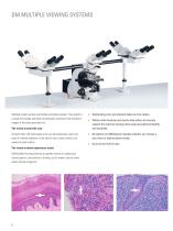
DM MULTIPLE VIEWING SYSTEMS Multiple header systems are flexible and highly modular. They attach to a single microscope and allow simultaneous viewing of high resolution images of the same specimen live. The vision to point the way A bright white LED illuminated arrow can be positioned to point out areas of interest anywhere in the field of view, clearly visible to all viewers at each station. The vision to make experience count DM Multiple Viewing Systems are perfect devices for obtaining a second opinion, consultation or training, as all viewers see the same superb sample image live. >...
Open the catalog to page 8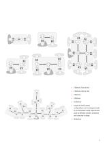
> 2 Stations, face to face > 2 Stations, side by side > 3 Stations > 5 Stations > 10 Stations > Large and small custom configurations can be designed easily to accommodate unique requirements such as different numbers of stations and room size or shape. > 20 Stations 491cm
Open the catalog to page 9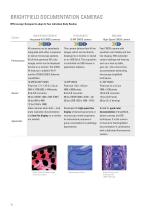
BRIGHTFIELD DOCUMENTATION CAMERAS MYcroscopy: Designed to adapt to Your individual Daily Routine IC90 E/ICC50 E/ICC50 W Integrated HD CMOS cameras HD BF DMC2900 High-Speed CMOS camera BF All cameras can be seamlessly integrated with either compound or stereo microscope systems. All of them generate HD color images, which can be displayed directly on a monitor. The ICC50 W features in addition Wi-Fi and the ICC50 E/IC90 E Ethernet capabilities. This camera delivers fast 4k live images, which can be directly displayed on a monitor or stored on an USB Stick. The acquisition is controlled via...
Open the catalog to page 10
Color CCD cameras High-Resolution CMOS camera Key success factors: > Leica color cameras provide state-of-the-art color interpolation algorithms performed in the camera head > Even fine structural and color details can be distinguished due to appropriate pixel sizes for every desired microscope magnification > High-Definition (HD) display directly on a monitor allows discussion of findings with a large auditorium Color camera High-Definition camera All contrast methods (except fluorescence) Dedicated fluorescence camera
Open the catalog to page 11
Pixel Shift Camera CCD Microscope Color Camera CCD Microscope Camera FLUORESCENCE DOCUMENTATION CAMERAS MYcroscopy: Designed with the highest sensitivity Cultured cortical neuronal cells (mouse).
Open the catalog to page 12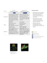
CCD Microscope Camera sCMOS Microscope Camera Key success factors: > High-sensitivity of the sensor allows short exposure times and therefore prevents photo bleaching and actively protects the cells from any photo damage > Cooling of the camera reduces unwanted noise and generates crystal clear fluorescence signals against dark background > Hardware-triggering and overlapping mode of read-out allows high-speed, real-time live cell imaging Color camera ^ Monochrome camera High-Definition camera All contrast methods (except fluorescence) >► Dedicated fluorescence camera
Open the catalog to page 13
OPTICAL BRILLIANCE The optical qualities of the DM microscope series are compelling. Outstanding image brilliance and razor-sharp contrast clearly reveal the most delicate specimen structures. The high level of comfort users expect contributes to fatigue-free viewing and greater efficiency. Quick changes between filter block positions. All filter blocks feature Zero Pixel Shift to prevent image shifting when superimposing different fluorescence excitations. Reduce eyestrain with HI PLAN SL Planachromat objectives. These objectives are designed to ensure the same level of brightness at all...
Open the catalog to page 14All Leica Microsystems catalogs and technical brochures
-
M844 F40/F20
16 Pages
-
EnFocus
12 Pages
-
M822 F40 / F20
12 Pages
-
M320 for ENT
12 Pages
-
ARveo 8
16 Pages
-
M320 Dental Brochure
12 Pages
-
Emspira 3
4 Pages
-
Exalta
2 Pages
-
FLEXACAM C1
4 Pages
-
EM KMR3
8 Pages
-
EM UC7
16 Pages
-
EM TRIM2
8 Pages
-
EM RAPID
8 Pages
-
EM ICE
12 Pages
-
EM TXP
10 Pages
-
EM RES102
12 Pages
-
EM TIC 3X
16 Pages
-
TL4000 BFDF
16 Pages
-
F12 I floor stand
6 Pages
-
XL Stand
4 Pages
-
TL3000 Ergo & TL5000 Ergo
4 Pages
-
KL300 LED
8 Pages
-
LED1000
16 Pages
-
LED3000 BLI
20 Pages
-
LED5000 NVI
20 Pages
-
LED3000 NVI
20 Pages
-
LED3000 DI
20 Pages
-
LED5000 HDI
20 Pages
-
LED5000 CXI
20 Pages
-
LED2500
8 Pages
-
LED5000 MCI
20 Pages
-
LED3000 MCI
20 Pages
-
LED5000 SLI
20 Pages
-
LED3000 SLI
20 Pages
-
LED2000
8 Pages
-
LED5000 RL
20 Pages
-
LED3000 RL
20 Pages
-
MZ10 F
4 Pages
-
M165 FC
16 Pages
-
M205 FCA, M205 FA
16 Pages
-
M125 C, M165 C, M205 C, M205 A
12 Pages
-
A60 F, A60 S
16 Pages
-
M50, M60, M80
12 Pages
-
DVM6
16 Pages
-
HCS A
20 Pages
-
TCS SPE
20 Pages
-
DFC450 C
6 Pages
-
DFC295
6 Pages
-
MC170 HD
6 Pages
-
DFC3000 G
6 Pages
-
DMC4500
4 Pages
-
ICC50 W, ICC50 E
6 Pages
-
DFC7000 T, DFC7000 GT
4 Pages
-
DFC9000
2 Pages
-
IC90 E
6 Pages
-
DMC6200
8 Pages
-
DMC5400
8 Pages
-
SFL7000
4 Pages
-
EL6000
4 Pages
-
SFL100
4 Pages
-
SFL4000
4 Pages
-
DMi8 S Platform
2 Pages
-
THUNDER Imager Live Cell
2 Pages
-
DMi8 M / C / A
12 Pages
-
DM IL LED
12 Pages
-
DMi1
6 Pages
-
DM3 XL
7 Pages
-
FS M
4 Pages
-
FS C
4 Pages
-
FS CB
4 Pages
-
DM3000, DM3000 LED
16 Pages
-
DM750 M
12 Pages
-
DM750
12 Pages
-
DM500
12 Pages
-
DM300
8 Pages
-
DM12000 M
8 Pages
-
DM8000 M
8 Pages
-
DM1750 M
12 Pages
-
DM4 M, DM6 M
12 Pages
-
DM4 P, DM2700 P, DM750 P
12 Pages
-
DM2000, DM2000 LED
16 Pages
-
DM1000
16 Pages
-
DCM8
16 Pages
-
DM1000 LED
16 Pages
-
DM4 B & DM6 B
16 Pages
-
DM6 M LIBS
2 Pages
-
S9 Series
12 Pages
-
Z6 APO
16 Pages
-
Z16 APO
16 Pages
-
Leica M530 OHX for ENT
4 Pages
-
M620 F20
8 Pages
-
M220 F12
8 Pages
-
Leica Application Suite X
4 Pages
-
Proveo 8
16 Pages
-
M525 F20
12 Pages
-
EnVisu Leica Handheld OCT
8 Pages
-
PROvido
8 Pages
-
Leica M530 OHX
16 Pages
-
Leica TCS SP8 STED 3X
24 Pages
-
Leica TCS SP8 Objective
24 Pages
-
Leica AOBS
16 Pages
-
Leica DMC2900
6 Pages
-
Leica DMshare
2 Pages
-
Leica_DMshare_ICC50-Flyer_en
2 Pages
-
Leica_DMshare_EC3-Flyer_en
2 Pages
-
Leica_SL801-Flyer
2 Pages
-
Leica_SCN400-Flyer_Clinical
2 Pages
-
DM2700 M
12 Pages
-
Leica_SR_GSD-Brochure
10 Pages
-
Leica_AF6000-Brochure
16 Pages
-
Leica motCorr-Flyer_EN
4 Pages
-
Leica TCS SP8-Flyer
2 Pages
-
Leica TCS SP8-Brochure
40 Pages
-
Leica TCS SP8 X-Flyer
2 Pages
-
Leica TCS SP8 STED-Flyer
2 Pages
-
Leica TCS SP8 HyD-Flyer
2 Pages


