- Catalogs
- Leica Microsystems
- EnFocus
- Company
- Products
- Catalogs
- News & Trends
- Exhibitions
EnFocus
1 /12Pages

EnFocus
1 /12Pages
Catalog excerpts

OCT systems for anterior & posterior segment surgeries FOCUS ON PERFECTION EnFocus intraoperative optical coherence tomography (OCT)
Open the catalog to page 1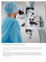
FOCUS ON PERFECTION Apply your skills with even greater confidence during eye surgeries with EnFocus intraoperative OCT built into the Proveo 8 ophthalmic microscope. Intraoperative OCT allows you to see what lies underneath the surface, giving you additional information for a complete understanding of how subsurface tissue reacts to your surgical maneuvers in real-time. At any step during surgery you can simply enhance your microscope view and add intraoperative OCT imaging with just a few taps. You get an immediate visual confirmation on ocular tissue behaviour so you can focus on achieving...
Open the catalog to page 2
“Having confirmation at every step during surgery is a huge advantage and helps enormously in surgical decisionmaking and diagnosis. In my experience intraoperative OCT makes the difference between compromise and perfection.” Dr. Barbara Parolini, Eyecare Clinic Brescia, Italy. Greater insight Supplement your microscope view with bright, sharp imaging of previously hidden subsurface details to better understand ocular pathology. EnFocus OCT imaging provides greater insights with additional information for your anterior and posterior segment procedures, you’ll see more at once. Immediate confirmation...
Open the catalog to page 3
GREATER INSIGHT Supplement your microscope view with bright, sharp imaging of previously hidden subsurface tissue details. EnFocus intraoperative OCT imaging provides additional information so you can get greater insights into ocular pathology during surgery. See hidden details with bright, sharp OCT imaging > Clearly differentiate between artifacts and tissue due to the unique spectrometer technology including dispersion compensation software and a highly sensitive detector that captures more signal > See fine details with an axial resolution of 2.4 μm in tissue due to the patented Leica spectrometer...
Open the catalog to page 4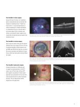
Your benefits in retina surgery Assess the level of tension in a membrane peel to avoid potential tears and protect the integrity of underlying tissue. A high-resolution view of ≤ 4 μm aids examination of retinal morphology for residual membranes or complications such as a macular hole or sub-retinal edema. Built-in dynamic scan control via footswitch further supports your visualization by aligning the scan angle to the membrane tissue. Microscope view of the retina (left) supplemented with EnFocus OCT (right) to visualize membrane layers during membrane peeling. OCT image courtesy of Seenu M....
Open the catalog to page 5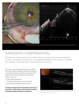
Measurement during corneal lamellar transplant surgery, courtesy of corneal lamellar transplant surgery, courtesy of Dr. Enrico Bertelli, head Bolzano Hospital, Italy. at Bolzano Hospital, Italy. Measurement during Dr. Enrico Bertelli, head of the ophthalmic department at of the ophthalmic department IMMEDIATE CONFIRMATION Confirm in real-time how ocular tissue is reacting intraoperatively to your surgical maneuvers. Immediately adjust your plan if needed for greater confidence in the surgical outcome. During retina, cornea and glaucoma procedures, there is typically a point at which surgeons...
Open the catalog to page 6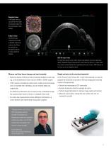
Surgical view In the quad view you can see the white light microscope image from the microscope’s video camera. Enface view The OCT B-scan composition provides a detailed anatomical surface view. Move the vertical line through the picture to review points of interest. OCT B-scan view The fast OCT scanner with a 30 Hz refresh rate delivers real-time subsurface details. Playback through acquired OCT scans frame by frame or in video mode for a careful review benefit from comprehensive scans of up to 1000 B-scans to not miss out on an important detail. Observe real-time tissue changes and react instantly...
Open the catalog to page 7
MAXIMUM FREEDOM EnFocus intraoperative OCT is built into your Proveo 8 and into your workflow. Switch view and record at the touch of a button with the confidence you will always have an optimal image. Our latest generation of EnFocus OCT was developed in collaboration with experienced ophthalmic surgeons for optimal workflow integration. No need to invest extra time and effort, or rely on a technician to activate OCT or to ensure you have the perfect image positioning and quality. Automatic image optimization and auto-locate functions do the job for you, with a single tap. You are free to concentrate...
Open the catalog to page 8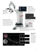
Choose how to view Inject OCT images directly into the eyepieces with the DI C800 module. Display your microscope and intraoperative OCT-image on the 27” touch-screen monitor. For even larger screen projections for you and your team, four video outputs are available. Switch your view with one tap Start surgery quickly Switch easily between the microscope view an intraoperative OCT view at any point yourself without interrupting surgery. Whether you use the footswitch, handle or the touchscreen monitor it’s just a single tap. Review acquired scans and recordings in the same way. Select, modify,...
Open the catalog to page 9
TECHNICAL SPECIFICATIONS PROVEO 8 WITH BUILT-IN ENFOCUS OCT Construction Floor stand Four 360° rotating castors (Ø150 mm), parking brake > Conforming with RoHS > Coated with antimicrobial paint > Floor stand max. 8.0 kg from microscope dovetail ring interface Direct illumination with 2 LED lamps > Floor stand approx. 380 kg without load, without built-in EnFocus OCT Main light > Integrated LED illumination system for intensive uniform illumination of the field of view > Continuously adjustable brightness with halogen-like color temperature CoAx 4 coaxial illumination > Illumination unit for generating...
Open the catalog to page 10
EnFocus (Ultra-HD) OCT Optical Performance > 15° /+ 105° motorized inclination tilt Axial resolution in tissue Zoom linked XY speed Lateral resolution Adjustable gas spring via balancing knob Floor stand with 4 electromagnetic brakes 860 mm flexible arm with 4 axis for rotation and inclination, max. weight 15 kg and up to 32” Control Control unit > User-friendly, individually programmable touch-screen (up to 30 surgeons) for control of motor functions and light intensity > Menu selection based on unique software for user-specific configuration > Built-in electronic auto-diagnosis and user support...
Open the catalog to page 11All Leica Microsystems catalogs and technical brochures
 M620 F20
8 Pages
 M844 F40/F20
16 Pages
 M822 F40 / F20
12 Pages
 M320 for ENT
12 Pages
 ARveo 8
16 Pages
 M320 Dental Brochure
12 Pages
 Emspira 3
4 Pages
 Exalta
2 Pages
 FLEXACAM C1
4 Pages
 EM KMR3
8 Pages
 EM UC7
16 Pages
 EM TRIM2
8 Pages
 EM RAPID
8 Pages
 EM ICE
12 Pages
 EM TXP
10 Pages
 EM RES102
12 Pages
 EM TIC 3X
16 Pages
 TL4000 BFDF
16 Pages
 F12 I floor stand
6 Pages
 XL Stand
4 Pages
 TL3000 Ergo & TL5000 Ergo
4 Pages
 KL300 LED
8 Pages
 LED1000
16 Pages
 LED3000 BLI
20 Pages
 LED5000 NVI
20 Pages
 LED3000 NVI
20 Pages
 LED3000 DI
20 Pages
 LED5000 HDI
20 Pages
 LED5000 CXI
20 Pages
 LED2500
8 Pages
 LED5000 MCI
20 Pages
 LED3000 MCI
20 Pages
 LED5000 SLI
20 Pages
 LED3000 SLI
20 Pages
 LED2000
8 Pages
 LED5000 RL
20 Pages
 LED3000 RL
20 Pages
 MZ10 F
4 Pages
 M165 FC
16 Pages
 M205 FCA, M205 FA
16 Pages
 M125 C, M165 C, M205 C, M205 A
12 Pages
 A60 F, A60 S
16 Pages
 M50, M60, M80
12 Pages
 DVM6
DVM616 Pages
 HCS A
HCS A20 Pages
 TCS SPE
20 Pages
 DFC450 C
6 Pages
 DFC295
6 Pages
 MC170 HD
6 Pages
 DFC3000 G
6 Pages
 DMC4500
4 Pages
 ICC50 W, ICC50 E
6 Pages
 DFC7000 T, DFC7000 GT
4 Pages
 DFC9000
2 Pages
 IC90 E
6 Pages
 DMC6200
8 Pages
 DMC5400
8 Pages
 SFL7000
4 Pages
 EL6000
4 Pages
 SFL100
4 Pages
 SFL4000
4 Pages
 DMi8 S Platform
2 Pages
 THUNDER Imager Live Cell
2 Pages
 DMi8 M / C / A
12 Pages
 DM IL LED
12 Pages
 DMi1
DMi16 Pages
 DM3 XL
7 Pages
 FS M
FS M4 Pages
 FS C
FS C4 Pages
 FS CB
FS CB4 Pages
 DM3000, DM3000 LED
16 Pages
 DM750 M
12 Pages
 DM750
DM75012 Pages
 DM500
DM50012 Pages
 DM300
DM3008 Pages
 DM12000 M
8 Pages
 DM8000 M
8 Pages
 DM1750 M
12 Pages
 DM4 M, DM6 M
12 Pages
 DM4 P, DM2700 P, DM750 P
12 Pages
 DM2000, DM2000 LED
16 Pages
 DM1000
16 Pages
 DCM8
DCM816 Pages
 DM1000 LED
16 Pages
 DM2500
16 Pages
 DM4 B & DM6 B
16 Pages
 DM6 M LIBS
2 Pages
 S9 Series
12 Pages
 Z6 APO
16 Pages
 Z16 APO
16 Pages
 Leica M530 OHX for ENT
4 Pages
 M220 F12
8 Pages
 Leica Application Suite X
4 Pages
 Proveo 8
16 Pages
 M525 F20
12 Pages
 EnVisu Leica Handheld OCT
8 Pages
 PROvido
8 Pages
 Leica M530 OHX
16 Pages
 Leica TCS SP8 STED 3X
24 Pages
 Leica TCS SP8 Objective
24 Pages
 Leica AOBS
16 Pages
 Leica DMC2900
6 Pages
 Leica DMshare
2 Pages
 Leica_DMshare_ICC50-Flyer_en
2 Pages
 Leica_DMshare_EC3-Flyer_en
2 Pages
 Leica_SL801-Flyer
2 Pages
 Leica_SCN400-Flyer_Clinical
2 Pages
 DM2700 M
12 Pages
 Leica_SR_GSD-Brochure
10 Pages
 Leica_AF6000-Brochure
16 Pages
 Leica motCorr-Flyer_EN
4 Pages
 Leica TCS SP8-Flyer
2 Pages
 Leica TCS SP8-Brochure
40 Pages
 Leica TCS SP8 X-Flyer
2 Pages
 Leica TCS SP8 STED-Flyer
2 Pages
 Leica TCS SP8 HyD-Flyer
2 Pages


- Leica analysis software
- Leica optical microscope
- Leica laboratory microscope
- Leica benchtop microscope
- Leica sample preparation system
- Leica visualization software
- Leica automatic sample preparation system
- Control software
- Leica LED microscope
- Leica laboratory software
- Windows software
- Leica laboratory sample preparation system
- Leica CMOS camera
- Leica camera with USB port
- Leica benchtop sample preparation system
- Automated software
- Leica LED light source
- Acquisition software
- Capture software