- Catalogs
- magtivio B.V.
- MagSi-STA 600, 1.0 and 3.0 L
MagSi-STA 600, 1.0 and 3.0 L
1 /2Pages
MagSi-STA 600, 1.0 and 3.0 L
1 /2Pages
Catalog excerpts

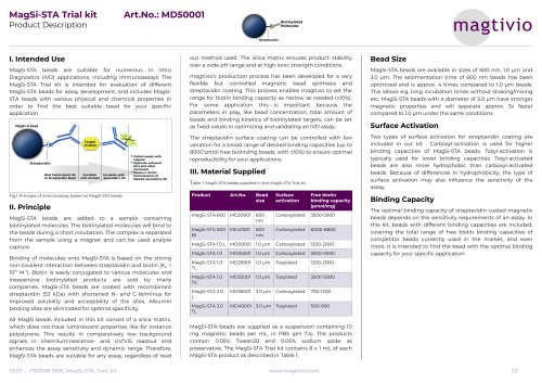
MagSi-STA 600, 1.0 and 3.0 L MD1X001, MD0X001, MD3X001 Product Description I. Intended Use MagSi-STA beads are suitable for many applications, including purification of proteins and peptides or nucleic acids, immunoprecipitation, immunoassays, protein interaction studies, phage display, and cell isolation. MagSi-STA beads are coated with streptavidin which efficiently binds to biotinylated molecules, e.g. peptides, proteins, antibodies, sugars, lectins. The magnetic properties enable easy and quick washing steps. Because beads are in suspension, incubation times can be shortened compared to alternative techniques. Columns or centrifugation steps are not necessary when working with magnetic beads. This feature enables easy implementation into automated processes. II. Principle MagSi-STA beads are added to a sample containing biotinylated molecules. The biotinylated molecules will bind to the beads during a short incubation. The complex is separated from the sample using a magnet and can be used in downstream applications. Binding of molecules onto MagSi-STA is based on the strong non-covalent interaction between streptavidin and biotin (Ka = 1015 M-1). Biotin is easily conjugated to various molecules and inexpensive biotinylated products are sold by many companies. MagSi-STA beads are coated with recombinant streptavidin (53 kDa) with shortened N- and C-terminus for improved solubility and accessibility of the sites. Albumin binding sites are eliminated for optimal specificity. MagSi-STA beads are available in sizes of 600 nm, 1.0 pm and 3.0 pm. The sedimentation time of 600 nm beads has been optimized and is approx. 4 times compared to 1.0 pm beads. This allows e.g. long incubation times without shaking/mixing etc. MagSi-STA beads with a diameter of 3.0 pm have stronger magnetic properties and will separate approx. 3x faster compared to 1.0 pm under the same conditions. III. Material Supplied 2, 10 or 100 mL MagSi-STA 600, MagSi-STA 1.0 or MagSi-STA 3.0 L. MagSi-STA beads are supplied at 10 mg/mL in PBS (pH 7.4), 0.05% Tween20, 0.05% sodium azide. Additional materials needed • Buffers and Materials (depending on the application, contact for support) • Magnetic separator for bead separation/collecting (see order information) • Mixer/vortex to homogenize samples and resuspend beads (depending on the application, contact for support) Washing and Binding Buffers • For coupling of proteins and peptides a neutral buffer (PBS) is recommended, optionally with a surfactant (0.05% Tween20) or 0.1% BSA to reduce background absorption. • For release of antigens from biotinylated antibodies, glycine 0.1M pH 2.8 (low pH elution) is recommended. Heating above 70°C in reducing SDS-PAGE buffer also releases antigens, but biotinylated antibody and streptavidin as well. • For binding of nucleic acids, TE-buffer (pH 7.5) is recommended IV. Product Use When stored at 2-8°C, this product is stable up to 2 years, but no longer than the expiry date on the label. Store in well closed vial and in upright position to prevent drying of the beads, this may result in a decrease of activity. Do not freeze the product! Vortex well before use. Wash the beads to remove preservatives that could interfere with your application. For washing, use the same volume as initially taken from the MagSi-STA vial or more. 1. Resuspend beads by shaking/vortexing 2. Pipette the required volume of beads into a tube or microplate (10-20 pL is suitable as a starting point) 3. Collect beads by placing the tube or microplate on the magnet for 1-2 minutes 4. While tube/micro plate is still on the magnet, carefully remove supernatant without touching the bead pellet 5. Take tube/micro plate from the magnet and add washing buffer 6. Resuspend beads by vortexing or pipetting 7. Repeat step 3 - 5 at least 3 times 8. Finally resuspend the beads in a suitable buffer for your downstream, in a volume equal to the original bead volume. 2. Incubate for 30 minutes at room temperature 3. Collect beads by placing the tube or microplate on the magnet for 1-2 minutes 4. Wash beads 3-4 times with washing buffer 5. Resuspend the beads in a suitable buffer and volume for your downstream use. 1. Combine the antigen sample with 10 pg of biotinylated antibody. Dilute each sample to a minimum volume of 300 pL with cell lysis buffer or Binding/Wash Buffer. Incubate 1-2 hours at room temperature or overnight at 4°C with mixing.
Open the catalog to page 1
MagSi-STA 600, 1.0 and 3.0 L MD1X001, MD0X001, MD3X001 Product Description 2. Resuspend beads by shaking/vortexing 3. Add 25-50 |jL of MagSi-STA beads into a 1.5 mL microcentrifuge tube. 4. Prepare the beads for binding by washing with binding buffer as described in a). Finally resuspend in 500 jL binding buffer. 5. Add the antigen sample/biotinylated antibody mixture to the 1.5 mL microcentrifuge tube containing pre-washed magnetic beads (4.) and incubate at RT for 30 minutes with mixing. 6. Collect beads by placing the tube on the magnet for 1-2 minutes, pipette off and save the supernatant...
Open the catalog to page 2All Magtivio B.V. catalogs and technical brochures
 rQ MagSi-NA Pathogens
2 Pages
 MM-Separator 50 P
2 Pages
 MM-Separator 32 FlipTube® BC
2 Pages
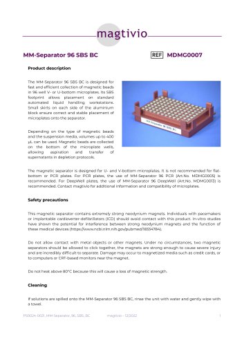 MM-Separator 96 SBS BC
2 Pages
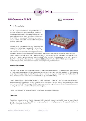 MM-Separator 96 PCR
2 Pages
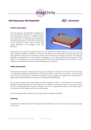 MM-Separator 96 DeepWell
2 Pages
 SafeQ Saliva Collection Kit
2 Pages
 MagSi-Tools 600, 1.0, 3.0
2 Pages
 MagSi-STA Trial kit
2 Pages
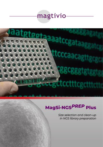 MagSi-NGSPREP Plus
2 Pages
 MagSi-DNA Stool
2 Pages
 MagSi-DNA FFPE
2 Pages
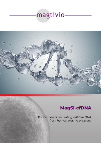 MagSi-cfDNA
2 Pages
 MagSi-DNA Tissue & Cells
2 Pages
 Product Catalog 2023
52 Pages

- Detection kit
- Solvent reagent
- Blood detection kit
- Molecular biology reagent
- Serum detection kit
- Plasma detection kit
- Research reagent
- Laboratory reagent
- Molecular detection kit
- Histology reagent
- Clinical detection kit
- Buffer solution reagent
- Immunoanalysis reagent kit
- Tissue reagent
- Urine detection kit
- DNA extraction reagent
- Laboratory assay kit
- Nasopharyngeal detection kit
- Magnetic bead-based reagent
- Collection kit