- Catalogs
- MDE Technologies
- Working Heart Perfusion setup
- Company
- Products
- Catalogs
- News & Trends
- Exhibitions
Working Heart Perfusion setup
1 /15Pages

Working Heart Perfusion setup
1 /15Pages
Catalog excerpts

heartperfusion Working Heart Perfusion setup
Open the catalog to page 1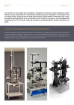
FOR OVER TWO DECADES, MDE HAS BEEN A PIONEER IN MANUFACTURING WORKING HEART APPARATUSES, CONSISTENTLY ADVANCING CARDIOVASCULAR RESEARCH. OUR DEDICATION TO EXCELLENCE IS REFLECTED IN OUR CONTINUOUS DEVELOPMENT PROCESS AND LONGSTANDING PARTNERSHIPS WITH ESTEEMED INSTITUTIONS, INCLUDING THE BUNDESWEHR TOXICOLOGY INSTITUTE AND VARIOUS ACADEMIC ESTABLISHMENTS ACROSS EASTERN EUROPE. A LEGACY OF COLLABORATION, INNOVATION, AND PRECISION Our latest collaboration with the Medical University of Vienna represents a significant milestone in MDE’s commitment to continuous development. Together, we proudly...
Open the catalog to page 2
UNVEILING PRECISION AND INSIGHT At the core of our approach lies the Working Heart In this model, the aorta of the heart can be connected via Method, emphasizing the maintenance of heart function a cannula to an aortic outflow line and initially perfused close to physiological conditions during perfusion. This in the Langendorff mode through a sidearm to the aortic methodology enables researchers to investigate critical line. A second cannula can be inserted into the left atri- aspects such as energy metabolism, oxygen consump- um, initiating heart function by clamping the retrograde tion, and...
Open the catalog to page 3
ADVANTAGES OF THE ISOLATED WORKING HEART MODEL IN CARDIOVASCULAR RESEARCH Since Neely’s pioneering work in 1967, the working heart model has garnered significant interest within the scientific community and has become a staple in laboratories worldwide. Broadly, there are two main types of isolated heart models: • The Langendorff isolated heart model (1895), where hearts receive coronary flow through retrograde perfusion. The working, fluid-ejecting heart model, in which hearts are perfused via the left atrium and eject fluid through the left ventricle into the aorta, thereby perfusing their...
Open the catalog to page 4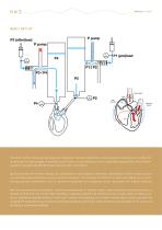
NEELY SET-UP The latter method, introduced by Neely and colleagues, has been adapted for various species, including mice. Notably, the working heart model engages in pressure-volume work, a crucial distinction from Langendorff preparations, which primarily perform energetically less demanding isovolumetric contractions. By perfusing the left ventricle directly, the working heart model allows for the direct measurement of left ventricular pressure and derivative parameters using a pressure transducer. This obviates the need for a ventricular balloon, thus circumventing potential confinements such...
Open the catalog to page 5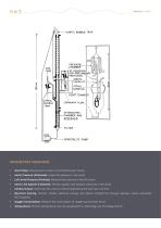
PARAMETERS MEASURED: • Heart Rate: Measure the number of contractions per minute. Aortic Pressure (Afterload): Assess the pressure in the aorta. Left Atrial Pressure (Preload): Measure the pressure in the left atrium. Aortic Line Systolic & Diastolic: Monitor systolic and diastolic pressures in the aorta. Cardiac Output: Determine the volume of blood ejected by the heart per unit time. Electrical Activity: Monitor cardiac electrical activity and detect arrhythmias through biphasic action potentials (ECG signals). Oxygen Consumption: Measure the consumption of oxygen by the heart tissue. Temperature:...
Open the catalog to page 6
RESEARCH FIELDS AND TYPICAL ISOLATED HEART STUDIES: Cardiovascular Physiology: Investigate fundamental mechanisms of cardiac contraction, relaxation, and electrophysiology. Study cardiac metabolism, systolic and diastolic dysfunction, myocardial function, and more. Pharmacology: Assess effects of drugs on cardiac function and electrophysiology, including ion channel blockers, inotropic agents, and antiarrhythmics. Conduct pharmacology/toxicology studies and ischemia-reperfusion studies. Disease Drug Development: Screen potential therapeutics for efficacy and safety in preclinical models. Test...
Open the catalog to page 7
MAIN FEATURES OF THE MDE ISOLATED WORKING HEART SETUP In addition to the basic setup, researchers have the option to enhance their experimental setup with supplementary parts and devices tailored to their specific requirements. These optional components include: Temperature Control: To ensure precise temperature regulation, each system includes a water-jacketed heart chamber. An additional heart chamber is available for non-invasive action potential measurement or creating a low-temperature environment for the heart. Switching between chambers is effortless due to their placement on suspending...
Open the catalog to page 8
Perfusion Modes: The apparatus operates in Langendorff constant pressure mode, with a roller pump supplying perfusate to the heart. In working-heart mode, the left atrium receives perfusate from the preload vessel through the atrial cannula. Pressure Regulation: To maintain constant pressure, the pump supplies more perfusate than required by the heart. Excess perfusate passes through a flow resistor back to the reservoir for oxygenation. The flow resistor can be adjusted to achieve the desired perfusion pressure. Seamless Mode Switching: The setup allows seamless switching between Langendorff...
Open the catalog to page 9
Drug Administration: Injection ports are provided at each cannula for the direct addition of drugs to the perfusate, enabling pharmacological studies.
Open the catalog to page 10
GENERAL CONSIDERATIONS FOR ISOLATED HEART PERFUSION STUDIES Typical composition of buffer for perfusion Procedure for salt mixture Krebs-Henseleit buffer Compound CARBOGEN GAS FOR OXYGEN SATURATION AND PH SETTING When preparing the buffer solution, solution has to be bubbled with Carbogen gas (95% O2 and 5% CO2) for at least 10 to 15 minutes prior to adding the final component CaCl2 to avoid precipitation of calcium as phosphate salt. Solubility (g gas/kg water)
Open the catalog to page 11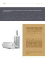
To avoid lack of oxygen and pH shift of the perfusion, the buffer solution has to be saturated with these gases. Saturation is temperature dependent. The plasma pO2 level between 100-120 mmHg results 100% haemoglobine saturation in blood. Red blood cells serve as O2 storage and when the plasma pO2 level decreases O2 diffusion from haemoglobine occurs to maintain the physiological plasma O2 level. Such storage is not available during buffer perfusion of the heart muscle therefore the buffer has to be saturated with O2. Proper saturation of buffer results in about 600 mmHg oxygen partial pressure...
Open the catalog to page 12All MDE Technologies catalogs and technical brochures
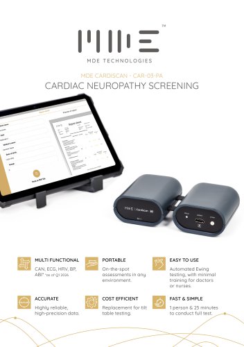 MDE CardiScan
2 Pages
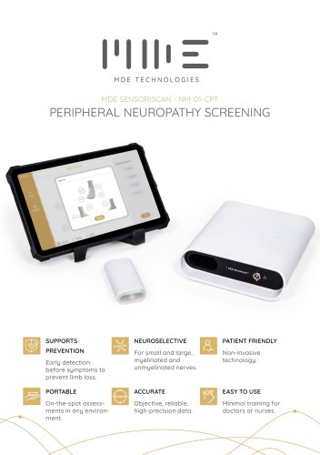 MDE SensoriScan -
2 Pages
 Heart Cell Isolation setup
10 Pages
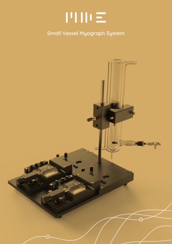 Wire Myograph System
22 Pages
 Langendorf_heart
14 Pages
 Isolated nerve tissue system
7 Pages
 Amplifiers and stimulators
9 Pages
 Cardiofish ECG device
16 Pages


- Laboratory bath
- Benchtop laboratory bath
- Vitals monitor
- Blood pressure vital signs monitor
- Circulating laboratory bath
- Intensive care vital signs monitor
- Compact vital signs monitor
- ECG vitals monitor
- Organ perfusion system
- Isolated organ perfusion system
- Heart organ perfusion system
- Animal research organ perfusion system
- Wearable vital signs monitor
- Small animal organ perfusion system
- Veterinary electrical stimulator
- Cardiology organ perfusion system
- Animal research veterinary electrical stimulator
- Organ bath
- Non-invasive vital signs monitor
- Myograph