MEDA/Fundus Camera/FDC-50A
1 /4Pages
MEDA/Fundus Camera/FDC-50A
1 /4Pages
Catalog excerpts

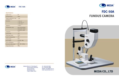
Specifications Angle of view: Working distance: Diopter compensation range for patient’s eye: Resolution: Brightness Working-table stroke: Vertical tilt: Lateral swing: Palate frame stroke: Light for photographing: Fixation lamp: Power Supply: Power consumption: Dimensions (Main body): Weight (Main body): CW) MEDA FDC-50A FUNDUS CAMERA Address:Room D, F3, Building C2, Xinmao Science Skill Park, Huayuan Industry Development Area, Tianjin, 300384, China
Open the catalog to page 1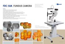
FDC-50A FUNDUS CAMERA Features: Colored, B&W, FFA and ICGA photography and comment; Convenient acquisition via handle, footswitch and mouse; Automatic identification between OS and OD; Dynamic angiography images displayed on the display screen in real time at 25 FPS; Full-information dynamic angiography images stored and cinelooped in real time at 25 FPS; Images analyzed, split/joined and processed on computer, and archived on DVD-RW; Images with adjustable brightness, contrast and color; Images denoised, smoothened, edge-enhanced and B/W converted on computer, length, area or PD measured on...
Open the catalog to page 2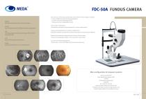
Display All fundus information from center to perimeter; Record Ever-changing fundus bloodflow throughout angiography; Edit Selected, analyzed and processed images and burn them onto a DVD-RW; Print Reports with images and comments; Replay Important segments for review and discussion. FDC-50A FUNDUS CAMERA The main unit ofthefundus camera take various kinds of fundus images, including: * Common B&W and colored fundus photography; * Fundus Fluorescein Angiography (FFA) * Indocyanine Green Angiography (ICGA) Various filter combinations: Automatically switched to address demand for various examinations; Unique...
Open the catalog to page 3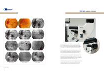
Gallery (for single case) FDC-50A FUNDUS CAMERA Optional: Facial Image Acquisition Unit Located under the main lens of the fundus camera, the facial image acquisition unit is uniquely designed for close-up microscopic HD photography that includes all capabilities on a slitlamp except for slit observation. Images, ranging from close-ups of lens and iris, to eye orbit, single eye, double eye, and furthertothe entire face, can be photographed by adjusting the scope. Corneal colored photography and iris angiography are also available using excited light emitted from the main lens of the fundus camera...
Open the catalog to page 4All MEDA catalogs and technical brochures
 MD-2300S AB Scan
2 Pages
 MEDA/Phaco/MD-480A
2 Pages
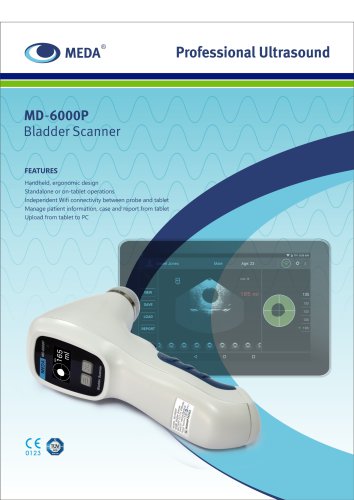 MD-6000P Bladder Scanner
2 Pages
 MD-960 Photocoagulator
2 Pages
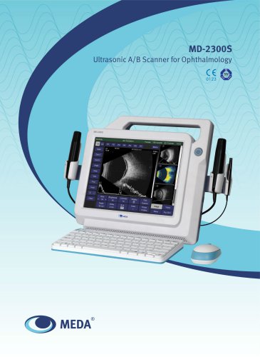 MD-2300S
4 Pages
Archived catalogs
 MEDA/A/B Scan/ODM-2200
2 Pages
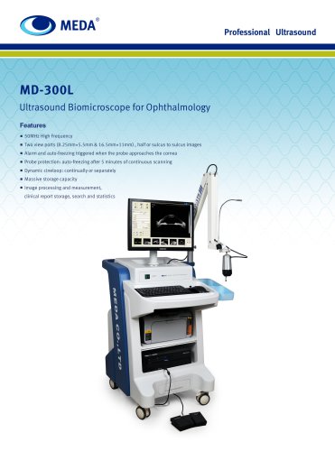 MEDA/UBM/MD-300L
2 Pages
 MEDA/A scan/MD-1000A
6 Pages
 MEDA/Auto Perimeter/MD-820A
2 Pages
 MEDA/skin scanner/MD-310S
2 Pages
 YAG Laser for Ophthalmology
2 Pages

- Med Tip ultrasound system
- Med Tip B/W ultrasound system
- Tabletop laser
- Portable ultrasound system
- Nd:YAG laser
- On-platform ultrasound system
- Med Tip fixed ophthalmic examination
- Touchscreen ultrasound system
- Hearing assessment system
- Nanosecond laser
- Ophthalmic laser
- Ultrasound bladder scanner
- Retinal photocoagulation laser
- Ophthalmic ultrasound imaging system
- Ophthalmic biometer
- Pachymeter
- Acoustic reflex tester
- Skin ultrasound imaging system
- Ophthalmic perimeter
- Capsulotomy laser