MEDA/skin scanner/MD-310S
1 /2Pages

MEDA/skin scanner/MD-310S
1 /2Pages
Catalog excerpts

• Display Window: • Display Mode: • Image Processing: • Database Management: CLINICAL APPLICATIONS • Specialized ultrasonic diagnostic system for dermatology application • Diagnoses of fine structures of epidermis, dermis, fine hair, subcutaneous tissue etc. • Observations of nevus, verruca vulgaris, scar, keratosis, scleroderma, hemangioma, skin tumors (benign tumors and cancers including lipomyoma, melanoma, basal cell carcinoma etc.) • Operation plan under instructions from ultrasonic diagnosis • Pathological observation during the drug treatment • Evaluation of laser treatment • Evaluation of skin condition - cosmetology • Non-invasive and comparable results. • 50MHz Very-High-Frequency Probe • Dermal Magnifier • Resolution up to 50 pm • Massive Image & Report Storage Capacity • Image Processing, Measurements, Retrieval and Printing 14mmxllmm & 7mmx5.5mm B&B+A Distance, area and angle measurements, color codes, y-correction and zooming Storage, Retrieval and Printing MD-310S Skin Scanner —High Frequency Ultrasound
Open the catalog to page 1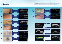
MD-310SSkin Scanner is applied to examination of fine structures of tissues like epidermis, dermis, fine hair and subcutaneous tissue. It can visualize the size and shape of kinds of nevus, scars, keratosis, scleroderma, hemangioma and skin tumors(including benign tumor or cancer such as lipomyoma, melanoma, basal cell carcinoma).
Open the catalog to page 2All MEDA catalogs and technical brochures
 MD-2300S AB Scan
2 Pages
 MEDA/Phaco/MD-480A
2 Pages
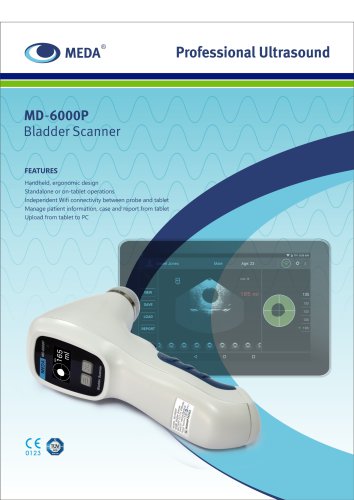 MD-6000P Bladder Scanner
2 Pages
 MD-960 Photocoagulator
2 Pages
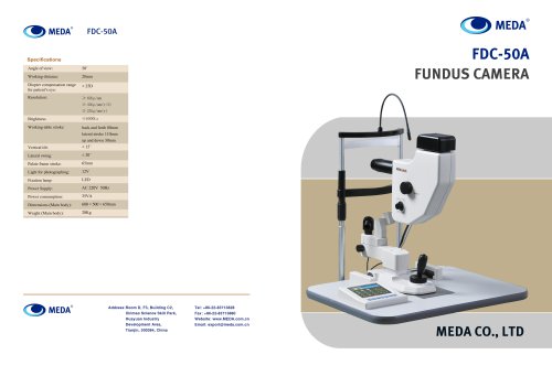 MEDA/Fundus Camera/FDC-50A
4 Pages
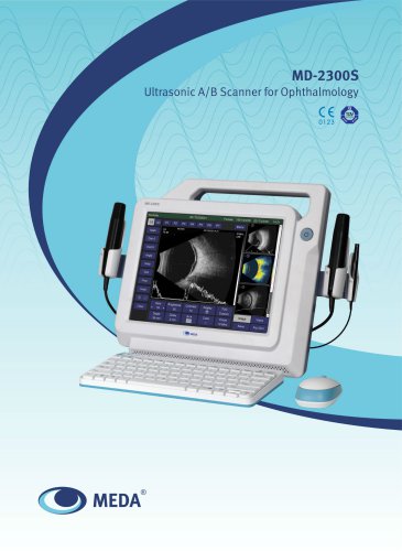 MD-2300S
4 Pages
Archived catalogs
 MEDA/A/B Scan/ODM-2200
2 Pages
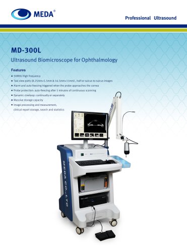 MEDA/UBM/MD-300L
2 Pages
 MEDA/A scan/MD-1000A
6 Pages
 MEDA/Auto Perimeter/MD-820A
2 Pages
 YAG Laser for Ophthalmology
2 Pages

- Med Tip ultrasound system
- Med Tip B/W ultrasound system
- Tabletop laser
- Portable ultrasound system
- Nd:YAG laser
- On-platform ultrasound system
- Med Tip fixed ophthalmic examination
- Touchscreen ultrasound system
- Hearing assessment system
- Nanosecond laser
- Ophthalmic laser
- Ultrasound bladder scanner
- Retinal photocoagulation laser
- Ophthalmic ultrasound imaging system
- Ophthalmic biometer
- Pachymeter
- Acoustic reflex tester
- Skin ultrasound imaging system
- Ophthalmic perimeter
- Capsulotomy laser