- Catalogs
- Meditech Equipment
- MD3203D
MD3203D

MD3203D
Catalog excerpts

MD3203D 3D ultrasound Scanner MD3203D is a high resolution 3D image(128 Elements)Specially designed keyboard greatly helps doctors from repetitive jobs. 3D Image processing algorithms include digital beam forming, multiply transmit focus point, adaptive aperture size, dynamic filtering, TGC, special and frequency mixing. 2 probe connectors can work at the same time. Features Literature Technical Gallery Main features Freehand 3D and Image Optimization software packages Full digital with latest ultrasonic imaging techniques and embed PC Portable design, easy for carrying Back-light, silicon-gel alphameric keyboard Applicable for abdomen, GYN, OB, cardiac, urology and small parts Supporting either video or laser printer 5 working modes: B, B/B, B/M, M and 4B 10¡± flash-free monitor, optional 10.4¡± medical LCD display 2 prob e sockets 8 segment TGC Permanent storage of 5,000 frames of ultrasound images, unlimited to S D card Peripheral ports of USB, VGA, video, DICOM3.0 and grounding Standard configurations main unit 3.5MHz convex probe with THI Probe holder Coupling gel
Open the catalog to page 1
Applicable Fields Suitable for the diagnosis of Abdomen, Cardiac, Gynecology, Obstetrics, Thyroid Gland, Small Organs, Urology and so on. W clinical examination and diagnosis. They are the ideal equipments to meet the needs of various kinds of hospitals and clinics. Technologies DBF: Digital Beam Forming RDA: Real-time dynamic aperture imaging DRA: Dynamic real-time acoustic apodizer DRF: D focus DFS: Dynamic frequency scanning, frequency range: 2.0-12.0MHz, 4 kinds of scaning frequencies Advantages: Embedded computer platform is adopted in the ultrasonic master system THI DFC Dynamic frequency...
Open the catalog to page 3All Meditech Equipment catalogs and technical brochures
 Oxima2 Vet
3 Pages
 Sonovet ID
1 Page
 BabySono
2 Pages
 SonoTech Pro
3 Pages
 SonoTech Lite
2 Pages
 SonoTech Xpress
2 Pages
 Sonotech M2
3 Pages
 Sono R
2 Pages
 iSono
iSono2 Pages
 Oxy plus
2 Pages
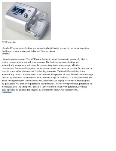 iBreathe CP
1 Page
 Defi6
Defi62 Pages
 FOs2
FOs22 Pages
 Spiro P
1 Page
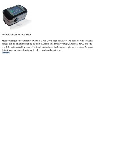 FOs3+
FOs3+2 Pages
 POxi2
POxi21 Page
 SpirOx Lite
1 Page
 SpirOx
3 Pages
 Micro convex probe
1 Page
 Transvaginal Probe
1 Page
 High freq Linear Probe
1 Page
 Convex Probe
1 Page
 MD3100
2 Pages
 MD8000
1 Page
 MD8015
2 Pages
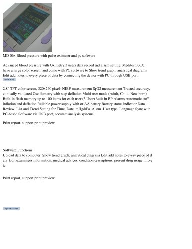 MD 06x
3 Pages
 MD-Touch
2 Pages
 MD 9015
1 Page
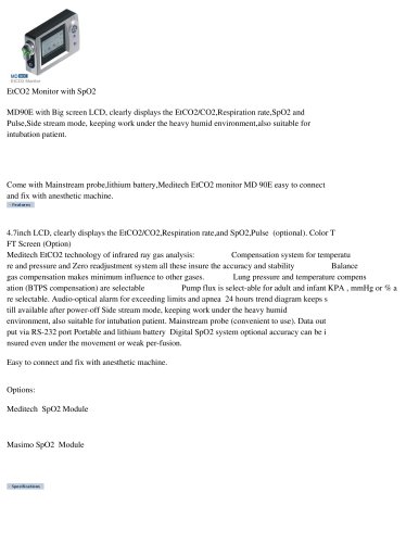 MD 90E capnography
2 Pages
 Echo80
2 Pages
 MDP100
1 Page
 ibeat
ibeat2 Pages
 EKG-6012
4 Pages
 EKG 1212T
2 Pages
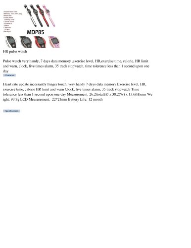 MDP85
MDP851 Page
 MDP90
MDP902 Pages
 MDP80w
1 Page
 Cardios pro
1 Page
 ECG-101T
2 Pages
 Defi
Defi2 Pages
- Logistics trolley
- Medical device trolley
- Ultrasound system
- B/W ultrasound system
- Blood pressure monitor
- Automatic blood pressure monitor
- Color doppler ultrasound system
- Patient monitor
- Portable ultrasound system
- Multipurpose ultrasound imaging system
- SpO2 monitor
- Blood pressure patient monitor
- ECG patient monitor
- EKG
- Digital electrocardiograph
- Ultrasound probe
- Veterinary ultrasound system
- Multipurpose veterinary ultrasound system
- SpO2 patient monitor
- Multi-parameter monitor