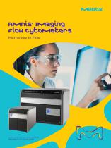
Amnis® Imaging Flow Cytometers

Amnis® Imaging Flow Cytometers
The document provides an overview of Amnis® Imaging Flow Cytometers, specifically the ImageStream®X MKII and FlowSight® systems. These instruments integrate the quantitative capabilities of flow cytometry with the detailed imagery of microscopy, serving various research fields such as immunology, microbiology, and oncology.
- FlowSight® Imaging Flow Cytometer: Utilizes camera-based detection for enhanced resolution and cost-effectiveness, enabling detailed cell population characterization.
- ImageStream®X Mark II: Offers high-throughput analysis with up to 60X magnification, user-friendly interface, and flexible laser configurations.
- Both systems provide multiple images per cell, including brightfield and up to 10 fluorescent markers, with high photonic sensitivity and minimal noise.
The systems are versatile, supporting research in:
- Immunophenotyping with multiple fluorescence channels for complex cell analysis.
- Quantifying nuclear translocation and cell signaling events.
- Studies on cell cycle, mitosis, DNA damage, and repair.
- INSPIRE® Software: Offers image-based gating, real-time fluorescence compensation, and intuitive sample handling.
- IDEAS® Software: Integrates image analysis with statistical rigor, providing comprehensive population statistics and flexible image display tools.
Amnis® Imaging Flow Cytometers provide a robust platform for cellular studies, delivering detailed imagery and quantitative data, suitable for both novice and expert users across various research disciplines.
- Dimensions: 889 mm x 660 mm x 635 mm
- Weight: 400 lbs (182 Kg)
- Illumination: Standard excitation at 488 nm; optional high power at various wavelengths. Side scatter at 785 nm standard. Brightfield is multichannel.
- Magnification options: 40x, 60x, 20x with respective numeric apertures of 0.75, 0.9, and 0.5.
- Pixel sizes: 0.5 x 0.5 µm, 0.3 x 0.3 µm, 1.0 x 1.0 µm.
- Field of view: 60 x 128 µm, 40 x 170 µm, 120 x 256 µm.
- Imaging rate: 2,000 cells/sec, 1,200 cells/sec, 4,000 cells/sec.
The system enhances performance through the Amnis® multispectral decomposition element, allowing simultaneous collection of brightfield, laser scatter, and multiple fluorescent images per cell.
The FlowSight® and ImageStream®X Imaging Flow Cytometers feature proprietary high-resolution cameras, optional second camera for 12 channels, standard and optional collection systems, filter wheels, lasers, and objective lenses. The system includes auto-focus, velocity detection, brightfield system, flow cell, laser retro system, and multi-spectral decomposition unit.
- FlowSight® Flow Cytometer: Catalog No. 100370
- ImageStream®X Mark II Flow Cytometer: Catalog No. 100220
- Reagents and Kits: SpeedBeads (400041), FlowSight® Calibration Beads (400300), Amnis® NFkB Translocation Kit (ACS10000), Amnis® Protein Aggregate and Silicone Oil Detection Kit (APH10001), Amnis® Intracellular Staining Kit (ACS10002).
For orders or technical assistance in Europe, contact customer service in respective countries or visit the Merck Millipore website for more information.
Catalog excerpts
Amnis Imaging Flow Cytometers ® The life science business of Merck operates as MilliporeSigma in the U.S. and Canada.
Open the catalog to page 1
Spanning the research disciplines in the life sciences Microscopy offers detailed cellular images and morphologic information, which are useful scientific tools for the study of cell function. However, the interpretation of microscopy images can be subjective, qualitative, and laborious. Flow cytometry is excellent for quantitative phenotyping and yields statistically robust results by rapidly interrogating large numbers of cells. However, flow cytometry lacks any ability to image, so sub-cellular localization and functional studies are difficult at best. By combining the speed, sensitivity,...
Open the catalog to page 2
Drug Discovery Stem Cell Biology Small Particle Analysis
Open the catalog to page 3
FlowSight® Imaging Flow Cytometer Capable: Applicable to every research discipline Sensitive: Camera-based detection dramatically increases resolution over traditional flow cytometry Affordable: Smaller footprint with configurations for any lab focus and budget Powerful: Characterizes populations by virtually any visual or fluorescent attribute
Open the catalog to page 4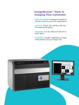
High-Throughput: Analyzes thousands of cells per second at up to 60X magnification Intuitive: Simple user interface with realtime plotting and gating Adaptable: Can be configured with one to seven lasers Boundless: Variable magnification images small particles and your largest cells
Open the catalog to page 5
powerful flow cytometry The ImageStream®X MKII and FlowSight® systems deliver multiple images of every cell in flow, including brightfield, darkfield (SSC) and up to 10 fluorescent markers at high speed. The ImageStream®X camera operates with a pixel size of 0.1/0.25/1 μm2 with 60X/40X/20X magnification, respectively, allowing visualization of fluorescence location from the membrane, cytoplasm, subcellular organelles or nucleus at high resolution. The FlowSight® system operates at 20X magnification with a 1 μm2 pixel. The innovative design of Amnis® cytometers increases signal and minimizes noise...
Open the catalog to page 6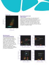
Single cells Beyond forward and side scatter Traditional flow cytometers do an admirable job of using low-resolution scattering characteristics to approximate size and intracellular granularity. Amnis® Imaging Flow Cytometers produce familiar 'size vs complexity' scatter plots, but with the power of 20x magnification—or more-can report absolute rather than relative cell size by measuring the actual diameter of objects in brightfield images. Multichannel Immunophenotyping Immunophenotyping requires multiple fluorescence channels in addition to dual scatter. Below is a six-color immunophenotype...
Open the catalog to page 7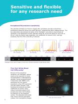
Sensitive and flexible for any research need Exceptional fluorescence sensitivity FITC Intensity The patented architecture of Amnis® imaging flow cytometers provides extraordinary fluorescence sensitivity across the visible spectrum, outperforming other imaging devices. The four plots below demonstrate the ability of the FlowSight® instrument to discriminate all intensities in the Spherotech 8-peak calibration bead set, across the spectrum from FITC to PE-Cy7. Note the distinct peak separation, low coefficients of variance (CVs) and high sensitivity from the FITC to the PE-Cy7 channels. FlowSight®...
Open the catalog to page 8
Images of every cell The FlowSight® and ImageStream®X instruments operate like conventional flow cytometers, but also provide imagery of every cell. Powerful and intuitive analysis software seamlessly links quantitative data to images: • Click on a dot in any plot to see its corresponding image • Click on a bin in any histogram to view every cell in that bin • Draw gates on dot plots and view the resulting populations to validate results With imaging capabilities, you'll never wonder about outliers or whether your gates are in the right place. Once you've drawn a gate on a plot you can click inside...
Open the catalog to page 9
Data acquisition software INSPIRE® software offers powerful image-based gating and real-time fluorescence compensation □ Instant Population Viewer Every population is added to a pull-down list as soon as you draw a gate. Simply select a population of interest from the list to view the corresponding cells during data acquisition. Image Gallery Imagery of cells of interest appear in the gallery as they are acquired, allowing you to inspect morphology, assess staining patterns, and optimize laser power settings. B Instrument Status at a Glance Convenient gauges, indicators, and text alerts provide...
Open the catalog to page 10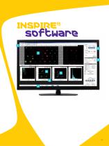
ElI< ** - h— > id^ # -b .■ ■ I ■ amnis ipriK M EW WH pill
Open the catalog to page 11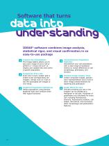
Software that turns data into understanding IDEAS® software combines image analysis, statistical rigor, and visual confirmation in an easy-to-use package 1 Images for Every Dot Every dot in every scatter plot is linked to the corresponding cell imagery. Simply click on a dot to see the associated cell images or vice-versa. Inspect Your Populations The Image Gallery allows you to see every image of every cell or perform a “virtual cell sort” to inspect and validate the cells within a specific population. Graphical Population Definitions Define populations using familiar graphical tools and combine...
Open the catalog to page 12
a wealth of applications Any Application You Can Imagine
Open the catalog to page 14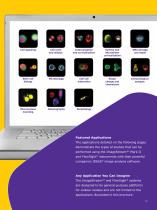
Cell signaling Cell cycle and mitosis Surface and intracellular co-localization Stem cell biology Cell-cell interaction Shape change and chemotaxis Immunological synapse Micronucleus Counting Featured Applications The applications detailed on the following pages demonstrate the types of studies that can be performed using the ImageStream®X Mark II and FlowSight® instruments with their powerful companion IDEAS® image analysis software. Any Application You Can Imagine The ImageStream®X and FlowSight® systems are designed to be general-purpose platforms for cellular studies and are not limited to...
Open the catalog to page 15
THP-1 Control (no LPS) Mean similarity score = 0.4 20X resolution tells the story Translocation of NFkB from the cytoplasm to the nucleus of the cell is a key event in the response to the presence of cell stressors. Only imaging flow cytometers can analyze translocation quantitatively, in thousands of cells. For this data, the 20x objective of the FlowSight® system is used to locate NFkB in relation to 7-AAD fluorescence from the nucleus in untreated THP-1 cells and cells stimulated with lipopolysaccharide (LPS). The similarity feature of the IDEAS® software produces a score for every cell quantifying...
Open the catalog to page 16All Merck catalogs and technical brochures
 Filters and Supporting Hardware
48 Pages


Archived catalogs
 Milli-Q HR 7000
12 Pages
 SDS 500
8 Pages
 Milli-Q® Integral system
22 Pages
 Elix® Advantage System
20 Pages
 AFS® 8D and 16D Systems
8 Pages
 ChromBook
406 Pages
 RiOs DI® Clinical
4 Pages
 Spectroquant® Prove
5 Pages
 Provantage® Services
2 Pages


- Detection kit
- Solvent reagent
- Blood detection kit
- Molecular biology reagent
- Serum detection kit
- Immunoassay detection kit
- Plasma detection kit
- Research reagent
- Diagnostic reagent
- Protein reagent
- Sample tube
- Optical test kit
- Laboratory test tube
- Clinical chemistry reagent kit
- Microtiter plate
- Medium reagent
- ELISA detection kit
- Antibody
- Gas filter
- 96-well microplate