- Catalogs
- Molecular Devices
- MetaFluor Software Brochure
- Products
- Catalogs
- News & Trends
- Exhibitions
MetaFluor Software Brochure
1 /6Pages

MetaFluor Software Brochure
1 /6Pages
Catalog excerpts
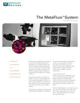
FLUORESCENCE RATIO IMAGING > RATIO IMAGING > CALCIUM IMAGING > FRET > pH MEASUREMENTS > ION CONCENTRATION > INTENSITY-OVER-TIME Fluorescence ratio imaging is the monitoring of live cells in which a fluorescent indicator of intracellular ions is introduced. Indicator dyes have been designed to shift their fluorescence excitation or emission spectrum when binding with specific ions. Images are obtained at two different wavelengths, typically matching the absorption bands at the high and low binding conditions. By ratioing the intensities in the images, it is possible to construct a map showing the local ion concentrations throughout the field of view. Since the monitoring process is nondestructive, image acquisition can be repeated frequently to trace and monitor the time course of cellular responses. The MetaFluor® Imaging System is designed for dual-wavelength intracellular ion measurements. The system provides simultaneous display of the raw data, ratio image, graphs of intensities, ratios and ion concentrations, and a nonratiometric image such as a brightfield or phasecontrast image. Two different ratiometric indicators can be imaged and measured simultaneously. CUSTOM CONFIGURATION Toolbars, menus, wizards and dialog boxes help move you through the image processing steps quickly. Features such as multiple image windows, flexible device control, synchronization and timing, and journals allow for automated image acquisition and analysis unlike any other system. With the MetaFluor System, you customize the set-up once, then let the experiment run by itself. You are able to collect a large amount of data online and process it with either MetaFluor or an analysisonly copy of the software.
Open the catalog to page 1
device control and system automation typical system configuration for fluorescence ratio imaging dichroic mirror light source filter wheel mirror DEVICE CONTROL MetaFluor works with microscopes equipped with epi-fluorescence illumination. The system includes device drivers for numerous commercially-available filter wheels, shutters, monochromators and high speed filter changers for illumination control. Camera drivers are optional. The MetaFluor system's camera drivers support acquisition from a wide variety of digital cameras. relay lens MetaFluor enables sub-region, binning and analog-to-digital...
Open the catalog to page 2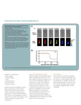
fluorescence ratio imaging applications Spatio-temporal activation of caspase revealed by indicator that is insensitive to environmental effects Kiwamu Takemoto 1,3, Takeharu Nagai 2,4, Atsushi Miyawaki 2, and Masayuki Miura 1 1 Laboratory for Cell Recovery Mechanisms and 2 Laboratory for Cell Function and Dynamics, Advanced Technology Development Center, RIKEN Brain Science Institute, Wako, Saitama 351-0198, Japan 3 Laboratories for Cell Biology and Neuroscience, Graduate School of Medicine, Osaka University, Suita, Osaka 565-0871, Japan 4 Structure and Function of Biomolecules, Precursory Research...
Open the catalog to page 3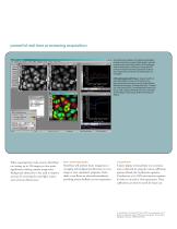
powerful real time processing acquisition A simultaneous display of multiple wavelengths images and various customizable graphs provide an easy point-and-click interface. When playing back an experiment, clicking on the graph will rewind or fast-forward the experiment to show the images that correspond to that location on the graph. CHO cells loaded with Fura-2. Experiment from the Optical Microscopy and Imaging in the Biomedical Sciences short course at the Marine Biological Laboratory, Woods Hole, MA. Courtesy of Lynda Pierini, PhD, Cornell Medical Center, Ken Dunn, PhD, Indiana University-Purdue...
Open the catalog to page 4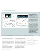
image analysis and processing In Vivo Imaging of the Dynamics of Glucose Uptake in the Cytosol of COS-7 Cells by Fluorescent Nanosensors Marcus Fehr, Sylvie Lalonde, Ida Lager, Michael W. Wolff, and Wolf B. Frommer The Zentrum für Molekular Biologie der Pflanzen Tübingen, Plant Physiology, Auf der Morgenstelle 1, D-72076 Tübingen, Germany Figure 3. In vivo characterization of FLIPglu-600µ. A, averaged YFP-CFP emission image shows FLIPglu-600µ in cytosol and exclusion from nuclei (N) and lysosomes (triangles). Emission intensities were higher in thicker layers of cytosol adjacent to the nucleus....
Open the catalog to page 5
METAFLUOR FLUORESCENCE RATIO IMAGING SYSTEM Technical Summary COMPUTER REQUIREMENTS DIGITAL CAMERA ACQUISITION FEATURES Intel® Pentium-4 processor or later > Microsoft® Windows® XP > CD-ROM drive > 512MB or more system memory (RAM) (more memory may be required for processing large image data sets) > 200MB free hard disk space for program only (image storage requires more space) > 24-bit graphics display (depends on imaging hardware used) > Exposure time, gain, A/D transfer speed, bitsper-pixel for each wavelength > On-chip gain multiplication > Binning and sub-region selection > Control of integrated...
Open the catalog to page 6All Molecular Devices catalogs and technical brochures
 ImageXpress Pico
8 Pages
 Threshold Immunoassay System
4 Pages


Archived catalogs
 Transfluor Technology
3 Pages
 CloneMatrix
2 Pages
 SpectraMax iD5
8 Pages
 GenePix 4000B Microarray
2 Pages
 EMax Plus Microplate Reader
4 Pages
 MetaMorph NX
4 Pages
 MetaXpress
6 Pages
 SpectraMax Paradigm
6 Pages
 SpectraMax i3x
6 Pages
 SpectraMax M series
6 Pages
 FlexStation 3
6 Pages
 SpectraMax MiniMax 300
3 Pages
 CloneMatrix - Data Sheet
1 Page
 EMax Plus Microplate Reader
3 Pages


- Detection kit
- Molecular biology reagent
- Laboratory reagent
- Analysis software
- Protein reagent
- Optical test kit
- Visualization software
- Microtiter plate
- Fluorescence detection kit
- Control software
- Medium reagent
- Antibody
- Reporting software
- Laboratory software
- Bacteria reagent
- 96-well microplate
- Research detection kit
- Monitoring software
- Planning software
- Cell assay kit