- Company
- Products
- Catalogs
- News & Trends
- Exhibitions
Mocean™ 4000
1 /8Pages

Mocean™ 4000
1 /8Pages
Catalog excerpts
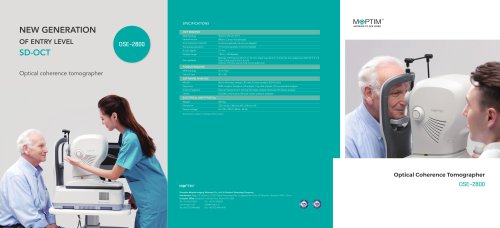
Optical Coherence Tomographer
Open the catalog to page 1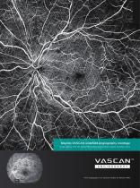
Moptim VASCAN widefield angiography montage Image courtesy of Dr. Bin Zhang, Peking University Shenzhen Hospital, Shenzhen, China * OCT angiography is an optional module for Mocean 4000
Open the catalog to page 2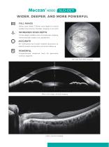
WIDER, DEEPER, AND MORE POWERFUL FULL RANGE 16mm scan width, 7.36mm scan depth (in tissue) enables the anterior chamber imaging in one shot INCREASED SCAN DEPTH 3.1mm depth enables clear choroid layer imaging, improving high myopic eye capabilities ACCURATE 45° SLO-based eye tracker enables physicians to identify lesions and perform accurate follow-up Comprehensive analytical tools for glaucoma, anterior segment 45° real-time SLO imaging 16mm full range corneal imaging
Open the catalog to page 3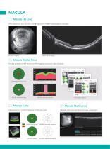
MACULA Macula HD Line High definition SLO and OCT imaging reveals hidden pathological changes Macula Radial Lines Have a glimpse of the retina via HD imaging and quick data analysis ILM/RPE thickness profile ILM/IPL thickness profile Macula Cube Macula Multi Lines Assessment of retinal thickness in 6x6 mm area ILM-RPE thickness ILM-RPE thickness deviation ILM-RPE volume Informative reports ILM-RPE volume deviation Multiple HD cross-sectional images acquisition
Open the catalog to page 4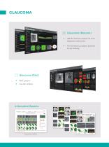
Glaucoma (Macular) • ILM-IPL thickness analysis for early diagnosis of glaucoma • Precise follow-up analysis powered by eye tracking Glaucoma (Disc) • RNFL analysis • Cup-disc analysis Informative Reports
Open the catalog to page 5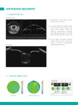
• Visualization of the entire anterior chamber (16 x 7.63 mm) • In standard mode, the system images from the corneal front surface to the lens's front surface, while the software automatically calculates ACD, ATA, pupil diameter, CCT, AOD 500, TISA 500, AOD 750, and TISA 750 • In lens mode, the system captures images from the front to the back surface of the lens, automatically measuring lens thickness
Open the catalog to page 6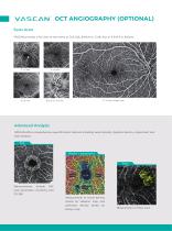
Scan Area VASCAN provides a full view of the retina at 3x3, 6x6, 8x8mm or 12x8, disc at 4.5x4.5 or 6x6mm. Advanced Analysis VASCAN offers comprehensive quantification features including vessel density, skeleton density, impairment and flow analysis. FAZ Density / Impairment area, perimeter, circularity, and FD 300 Measurement of vessel density based on skeleton map and perfusion binary map Measurement of flow area
Open the catalog to page 7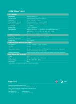
Methodology Spectral domain OCT Optical source Superuminescent diode (SLD), 840 nm Scan speed 80,000 / 120,000 A-scans/s Axial resolution (optical) 5 microns (optical), 3.6 microns (digital) Transverse resolution 15 microns (optical), 3 microns (digital) Diopter range - 20 to + 20 diopters Scan patterns Macular: HD line (6 / 12 / 16mm), 3D (6 x 6mm), 6 radial lines Multi lines (X-Y: 5 x 5 / X:10 / Y:10); Disc: 3D (6 x 6mm) Anterior: HD line scan (6 / 16mm), 6 radial lines Methodology Line scanning laser ophthalmoscopy (LSLO) Minimum pupil diameter 3.0 mm Field of view 45 ± 1 degrees Algorithm...
Open the catalog to page 8


Archived catalogs
 OSE-2800
2 Pages
 iRef
iRef3 Pages
 Mocean 4000
8 Pages
 Colombo IOL Optical Biometer
3 Pages
 MOPTIM catalogue 2022
12 Pages
- Fixed ophthalmic examination
- Software module
- Laboratory refractometer
- Hand-held ophthalmic examination instrument
- Slit lamp
- Digital refractometer
- Table slit lamp
- Ophthalmoscope
- Operating microscope
- Portable refractometer
- Analysis software module
- Refractometer ophthalmic examination
- Automatic refractometer
- Medical imaging software module
- Ophthalmic biometer
- Visualization software module
- OCT ophthalmoscope
- Tablet PC software module
- Retinal imaging instrument
- Dry eye diagnosis system